How to use canine splints

Canine splints are the new contribution to the treatment of lesions in the distal limbs of dogs.

Splints are useful both in neurological problems, where they position the dog’s foot or hand well; as in traumatological problems where they support and immobilize.

Before using a splint it is important that we make sure that the size is correct, a small splint would compress too much and a very wide one would dance and not give a good support, being able to generate chafing. Splints can be trimmed in some cases where necessary but the angle of the natural position of the limb should not be modified.

Splint placement

The first few times the splint is placed, it is better to have the help of another person who holds or distracts the dog.

We can put the splint of the anterior limb with the dog sitting or with the dog in station (standing with all four limbs on the ground; it is not advisable to put the splint with the dog lying down neither in lateral decubitus nor in ventral decubitus.

250x250-2

The splints of the posterior ones should be placed with the dog in season.

Of course, the dog’s skin should be clean and dry, it is very important that the hair is dry since otherwise it can cause skin irritations, redness or small ulcers.

It is advisable to put a tubular bandage or a cohesive elastic bandage around the dog’s paw. The function of this bandage is to increase comfort, it should not be too tight and will make the dog tolerate the splint better.

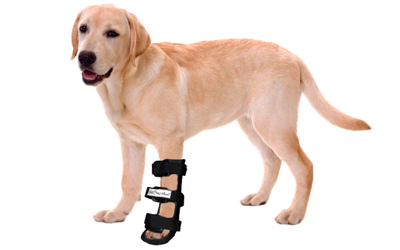
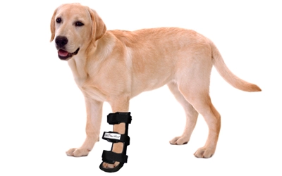
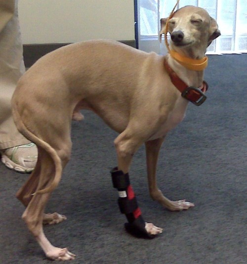
The splint is placed on the back of the forelimb leaving the Velcro straps on the front, first place the dog’s pads on the bottom of the splint, leaving the fingers relaxed, these should protrude from the splint.

Once put in situation, the protectors of the front of the straps will have to be right in the middle of the front of the dog’s leg for greater comfort. Adjust the lower Velcro first making sure that the dog’s leg is fully adjusted to the splint, then adjust the middle strap and finally the upper one.

Inmovilizador pata perro

Unless instructed by your veterinarian should not wear the splint throughout the day, normally at night the dog can sleep without it, and during the day we will put it on especially in the moments of more exercise. In certain neurological problems or after fissures or fractures, according to the advice of your veterinarian, it may be indicated to wear it permanently. In the latter case we must regularly monitor the skin of the animal.

Its use is solving cases in which traditional immobilization had failed and many animals can regain their mobility thanks to the invention. We hope that in the future it will help many more dogs.


Orthocanis Team

Baner silla 728x90

Elbow dysplasia in dogs

Elbow dysplasia is a very common degenerative disease in young dogs.

The elbow of dogs is one of the most congruent and stable joints of the body, allowing, due to its complexity, two axes of or degrees of supination-pronation movement of the forearm and flexion-extension. Its complexity is given by its composition: humeroradial joint, humeroulnar and, proximal radioulnar.

Elbow dysplasia was initially used to describe the non-union of the anconeal process (AUP). Currently, osteochondritis dissecans (OCD) of the medial condyle of the humerus, the fragment of the coronoid process (FPC) and, the incongruence of the elbow (INC) are also included within this term. When one of these ossification defects occurs in an elbow, inflammation originates and over time an osteoarthritis is triggered in which cartilage degeneration occurs; for that reason, all these conditions are commonly associated with osteoathrosis of this joint and are an important cause of pain and claudication of the forelimbs in large and giant breed dogs such as the German Shepherd, Labrador, St. Bernard, Rottweiler, Neapolitan Mastiff, among others.
protector-codo-canino

Of multifactorial genetic origin, especially in OCD and FPC. It affects males more than females and can occur uni- or bilaterally. The genetic component is the one that has the greatest influence although, the appearance of this pathology can also occur due to food, weight, environment, quality of ligaments, a lot of physical exercise or trauma.

The first symptoms may occur at 4-5 months when the dog shows exercise intolerance, lameness when starting a movement or after prolonged exercise. There are dogs that do not show signs of affection in the elbow until advanced ages where the process of osteoarthritis is very evolved. Others manage to maintain a normal degree of activity throughout their lives.

The fact of making a premature radiological diagnosis makes it possible to establish an adequate treatment and avoids the formation of osteoarthritis that produces pain and functional limitation of the elbow throughout the life of the animal. The diagnosis can be complemented with diagnostic tests such as CT or MRI

The evolution depends on the degree and type of injury, but it is usually unfavorable without surgery. Surgical treatment is good if degenerative changes in the joint have not yet occurred. In any case it is necessary to perform a good rehabilitation in order to:

  • Speed up the recovery process
  • Eliminate pain and inflammation
  • Decrease lameness
  • Maintain and/or improve range of motion
  • Maintain muscle tone, mass and strength
  • Minimize or slow down the effects of joint degeneration – osteoarthritis
  • Avoid compensation at the level of the neck, spine and extremities
  • Give the maximum capacities so that the animal is functional and that it, with a good quality of life

Physiotherapy treatment varies depending on the animal and the state of the lesion. It is important to start as soon as possible with the treatment so that it is effective and, to avoid drying them as reduced mobility and / or chronic pain.

The animal goes through different phases until its full recovery. It is essential to gradually achieve the objectives set. The recovery process is terminated when the animal is able to perform daily activities.

During the first three days after the intervention, it is important to act on inflammation and pain and prevent muscle atrophy and decrease in the joint arch from appearing. For this, passive techniques are used that reduce inflammation, produce analgesia and help maintain tone, mass and the arc of mobility. Among these techniques there are electrotherapy (segmental TENS and muscle electrostimulation), massage, passive mobilizations and cryotherapy (cold).

In older dogs or dogs that have not been intervened, the objectives will be the same as in animals that have gone through an intervention. It is important to eliminate pain because, with pain you can not work.

It is important from the beginning to massage and move the affected elbow as long as there is no veterinary contraindication and, respecting in the case of fixation, the period of healing and union of the fixed parts. Massaging and moving the affected area and limb helps maintain mobility, prevents loss of mass and tone and works the proprioceptors.
ortesis-codoA gentle mobilization combined with different massage techniques help decrease inflammation and reduce pain.

With TENSat the segmental level we can produce analgesia and decrease the amount of drugs administered. There are animals that have intolerance to certain drugs that produce analgesia and with TENS the pain can be reduced. TenS can also be used directly on the injured or operated area, as long as there is no osteosynthesis material underneath, since an internal burn could occur.

Muscle electrostimulationhelps prevent the onset of atrophy and maintain muscle mass and tone. With electrical stimuli we can stimulate nerve conduction.

At the beginning and end of the session the coldis used since it has properties that act on the decrease of the inflammatory response, edema and pain.

From the fourth day and during the next two weeks when the inflammation and pain have disappeared it is time to introduce simple active exercises such as shaking hands or small walks on a leash to force the animal to make an equal support with the four limbs and thus, prevent a decompensation between limbs from appearing due to not having a correct support on the ground. The walks is an exercise that increases the duration until full recovery.

Once the stitches have been removed, the animal can be introduced into the water. The advantages of water are used to improve recovery. Hydrotherapy (underwater treadmill) facilitates the station of the animal without loss of balance and, thanks to flotation, without having to support all its weight. In addition, flotation allows animals with bone pain and low muscle mass to work. The pressure of the water exerted on the body of the animal increases the sensitivity and decreases inflammations and edemas. The work in the water, underwater tapes or swimming increasesas the animal recovers. In addition, with water, we can recover the motor pattern, increase mass, tone and strength, work on respiratory capacity and maintain and / or improve mobility.

Once the acute phase has passed 48-72 hours and without risk of infection or inflammation, heat can be introduced that helps to elastify the tissues, decreases pain and increases vascularization among others.

The use of boards, plates, balls and trampolines are important to work on balance, proprioception and above all the integration of the affected limb.

It is already in the last phase, from two weeks, when the dog has integrated the gait pattern, exercises are performed to improve the quality of movement. They are more complex active exercises to integrate the affected limb or limbs. With active and proprioception exercises it is possible to increase muscle tone, mass and strength; coordination and balance and range of motion are worked on. Rails with different surfaces, cones, bars, circuits, up and down stairs and ramps (staircase with inclined plane) are used.

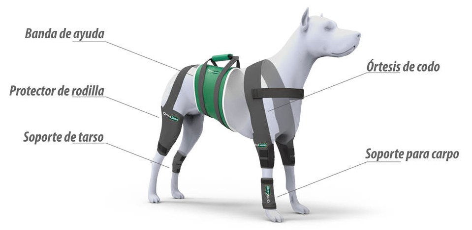
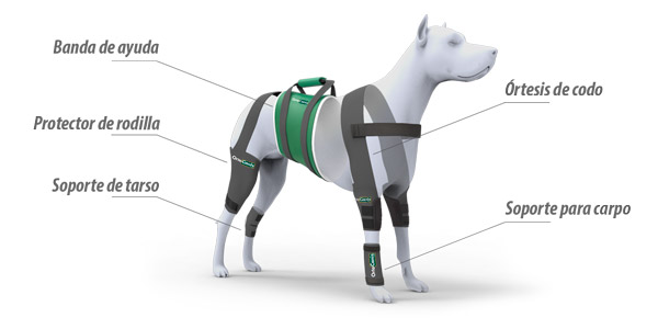
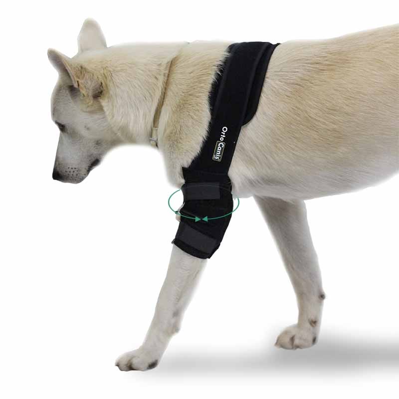
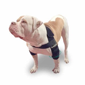
Throughout the recovery treatment and in animals with developed osteoarthritis it is essential to reduce the weight on the joints of the elbows. For this purpose , special support harnesses for elbows are used. In addition to decreasing weight, pain is reduced and does not hinder movement, the animal feels more comfortable; the joint is protected at all times from chafing and blows and helps maintain the heat that the animal gives off, which leads to a relief of the affected area.

At home, special care should be taken for animals suffering from elbow conditions. This care is necessary during and after treatment:

  • Avoid slippery floors
  • Avoid ramps and stairs at the beginning of treatment in operated animals and in animals that do conservative treatment. Once rehabilitated, ramps can be used to help get on the sofa and the car, since it is recommended that they do not do it alone, there could be a recidivism.
  • It is recommended that they rest on soft and clean surfaces, but that they are firm enough to help the incorporation of the animal special mattress for dogs
  • Keep skin clean and dry skin
  • Use special plates at your height so as not to strain the elbow joints
  • Correct diet and weight control. Being overweight harms the joints and generates more pain for the animal

It is very important to create an exercise routine and environment to help keep the animal comfortable and with quality of life.


You can also check:

Can chondroprotectors help?

Elbow dysplasia in dogs

Hip dysplasia in dogs

Baner silla 728x90

Cold weather and osteoarthritis in the dog

Osteoarthritis is a very common degenerative disease of the joints in dogs. Osteoarthritis causes pain, decreased joint range of motion, and joint inflammation.

The cold weather and especially the humidity, can increase the symptoms of this pathology, in autumn and winter is when dogs with osteoarthritis suffer the most.

Two types of osteoarthritis are distinguished, primary and secondary. The primary ones are degenerative, can affect more than one joint and are due to age and the “wear” of the joint. They are the typical osteoarthritis of the knee, carpus, tarsus and even spine that the elderly human population also suffers. Secondary osteoarthritis is due to joint misalignment that has prematurely worn down the articular cartilage. These occur after a fracture, especially if it has affected the joint, due to a bad joint disposition (demarcation): bad aplombs, or in the most common house secondary to hip dysplasia.

perro-multimedia-600x300_6

Photo: different joint protectors for dogs

Both in one case and in the other several things are recommended:

  • Strict control of the diet: the dog must be at its ideal weight, if it is overweight the joints suffer very significantly this excess.
  • Anti-inflammatory medication, now recommended NSAIDs cox-2 are new generation nonsteroidal anti-inflammatory drugs with far fewer side effects and more direct action on areas of pain.
  • Joint protectors: glycosaminoglycans, chondroitin sulfate…
  • Regular physical exercise: it is very important not to lose too much muscle mass, dogs with osteoarthritis usually weaken the muscles due to lack of use, this weakening and muscle atrophy worsens the picture
  • Sleep in padded beds, insulated from moisture and warm.
  • Joint protectors: supports to respect joint function.
  • Avoid exposure to cold and sudden changes in temperature: blankets to protect them from the cold and humidity can help us.

There are specific products to protect and support the joints of our animals, both for the protection of the tarsus and for the carpus.

You can also find blankets or coats for dogs that reflect the heat of the same animal and help in cases of osteoarthritis of the spine and hip. And special mattresses for the relief of the ailments of osteoarthritis in the dog. In the morning when the affected joint is cold and the animal has not moved for a long time, the symptoms will be more evident.

Back on Track products are made in Sweden and are made with so-called “smart textiles” or new generation unique textiles that have been developed on the basis of knowledge of ancient Chinese medicine along with the most modern research, as for the techniques applied to the textile industry, supported with scientific studies. The resulting product has been a fabric formed by an optimal fusion of polyester/polypropylene and ceramic particles.

Ceramics reflect body heat by restoring it in the form of infrared radiation. It is well known that infrared light has a calming effect as mild heat reduces inflammation, decreases muscle tension and improves blood circulation. The muscles in tension relax and the process of muscle recovery is accelerated, tendons, ligaments and joints injured and sore.

01_perrocorriendonieveabrigo
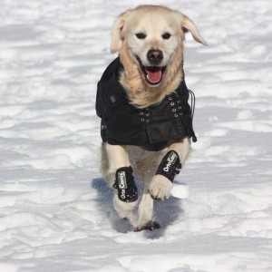
Photo: Thermal coat, protects from cold and humidity conserving the heat of the animal itself.

The essential function of tissue with ceramic particles is to prevent damage, as well as relieve and accelerate the process of recovery from injuries but it is also used to warm the muscles before exercise or physical work, thus eliminating the risks of pulls and fibrillar tears.

These products: Increase blood circulation, accelerate recovery from injuries, reduce inflammation, reduce muscle tension and relieve pain.

Fractures in dogs, how to act?

Source: Eroski Consumer

Without knowledge of first aid, the dog’s broken limb should not be moved so as not to injure the area further.

Inmovilizador pata perroAccidents and falls are the most common causes of fractures or broken bones in dogs . But not all fractures are the same. When they are serious and the dog does not receive treatment, it can die. This article explains how the owner of a dog with a fracture should act , how to prevent them and which dogs are more at risk of suffering fractures , as well as the reasons.

How should the owner of a dog with a fracture act?

If you have some knowledge of first aid, you can immobilize the injured area with a magazine or newspaper.

Owners can follow certain guidelines when their dog sustains a fracture . The main one is to go to the vet as soon as possible and move the dog as little as possible during its transfer. If you have some knowledge of first aid, you can immobilize the injured area with a magazine or newspaper tied or bandaged around the affected limb.

If you don’t know how to do it, because you don’t have minimal knowledge about how to splint the limb, it is recommended not to manipulate it. There is a risk of further injuring the area. Fractures located below the elbow or knee, in the joints, are more likely to worsen if the dog is moved.

Dogs can become unconscious after sustaining a fracture . In these cases, it is advisable to move them with their heads raised and not bend or compress the neck. In this way, we will prevent them from swallowing their tongue and they will be able to breathe. In addition, the danger of injury to the cervical area is reduced.

The speed with which we move the dog to the vet is essential. It may be that the accident occurs during the weekend or on a holiday, but there are veterinary clinics that have an emergency service and are available 24 hours a day.

Some of these clinics have an ambulance for the urgent collection of injured animals. Keep in mind that internal injuries can be more serious than fractures, so the sooner they are located and treated, the better.

“Fractures that occur in flat bones heal by themselves,” says veterinarian Ángel Suela. “However, those located in long bones, such as the femur, which break the tissue, are more serious fractures,” he says. There are those that may need surgery or cards (similar to a plaster) that immobilize the affected area for a month.

How to prevent bone breaks in the dog

Falls and accidents are frequent reasons for traumatism in the dog.

The best way to prevent fractures in dogs is to avoid accidents that cause trauma. “The gutters, drains and sewers are places where the dog suffers fractures and accidents are frequent,” warns veterinarian Leire Jiménez. Most dog fractures are due to falls and run overs . “There are dogs that fall out of the window or put their foot in a place where it gets caught and breaks,” he explains.

Solución para fractura perroIf you have a garden at home, make sure there are no places where the animal can get injured. Any gaps where their legs get caught should be plugged. If it is a dog that likes to look out the window -and is nervous and active, like a puppy-, it is advisable to close the windows or place parapets that prevent it from falling.

Hit-and-runs are also a source of fractures and injuries, which can be life-threatening. Therefore, the animal must circulate on a leash on public roads. In the countryside, although the dog walks without a leash in some sections, it must be supervised by its owners. The animal may escape and be run over on a nearby road or fall down a ravine or well.

Dogs at higher risk of fractures

Fractures or broken bones are more likely to occur in puppies, as they are very restless and are more at risk of falls and accidents. But dogs that suffer from bone wear (osteoporosis) or are very old are also candidates.

  • Very old dogs, from 10 years. They are more at risk of breaking a bone and their fractures take longer to heal. Lack of calcium, due to age, slows down fracture repair.
  • Dogs suffering from osteoporosis or decalcification of the bones. They are more at risk of fractures because their bones are weaker and a blow can cause them to break.
  • Puppies and very active dogs. Above all, when the dog has access to a garden, it should be watched, since it is more at risk of falling and hitting.


Caroline Pinedo

Source: Consumer

Degenerative myelopathy, the progressive and degenerative disease of the spinal cord of the elderly dog, has an onset from 8 years of age. In the initial phases, the dog shows incoordination in movements, falls off its hind legs or makes strange movements; staggers and drags one or both feet or walks with knuckles.

The disease can start in one hind limb and affect the other up to the thoracic limbs. The weakness progressively worsens, the dog has difficulty standing and has trouble walking. The disease can progress for a year until the dog finally becomes paraplegic or tetraplegic and must be euthanized. It is a slightly painful disease and in most cases it affects urination and defecation, they become incontinent.

There are no treatments that reduce this degeneration or stop it, but there are measures to help these dogs that suffer from it to maintain their quality of life. It is necessary for the animal to do rehabilitation exercises and maintain lifestyle habits at home to avoid pressure ulcers, urine infections and loss of mobility.

Physiotherapy and rehabilitation can help slow the process. Secondary pain symptoms (tensions), created by the animal itself when trying to move, can be controlled. The aim is to stop the appearance of atrophy (loss of muscle mass) and preserve the function of the forelimbs, as well as preserve the integrity of the hind limbs, to avoid ulcers and keep them active as long as possible, stimulate their sensitivity and work on coordination and balance of the animal to give it a better quality of life.

For this, passive mobilization exercises, massages, stretching, combined with heat, hot packs , and with some device that combat atrophy , muscle electrostimulators and TENS pain, are used.

arnes-de-soporte-para-perro

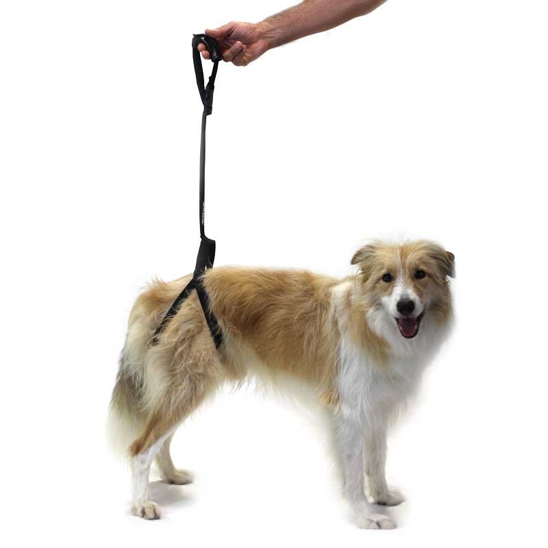
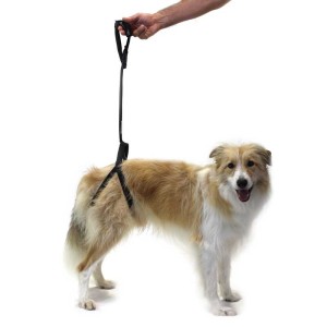
There is another part of the therapy that tries to maintain active mobility on the part of the animal. For that, balls , plates , hydrotherapy, bars and cones , walks, etc. are used. to maintain active mobility with good coordination without loss of balance. This stage is the one that is most altered with the course of the disease and for this, when the animal begins to deteriorate, not maintain its own weight, it is necessary to maintain mobility with the use of Harnesses for hindquarters , in the case of presenting difficulty only in hindquarters, or integral harnesses , to also support the anterior limbs.

The latest novelty on the market is the Biko Brace , a device that allows the dog to walk when the disease already affects the hind limbs significantly. In advanced phases of the disease, the use of customized wheelchairs is necessary.

During the stay at home, the animal must be in a comfortable, soft but firm place so that it can be easily incorporated. special mattress for dogs If the disease is in its early stages and the animal is dragging its feet, or with its knuckles, it is important to protect that area to avoid ulcers with booties or socks for dogs

During the course of the disease, the nutrition and weight control of the animal is very important to avoid complications.

In the following video we see TEX, a German Shepherd affected by degenerative myelopathy before and after the use of the Biko-Brace device.

Marta Subirats

canine physical therapist

Certified Canine Rehabilitation Practitioner by the University of Tennessee

Advice after surgery

All surgical intervention entails certain problems of pain, osteoarthritis, mobility, loss of muscle mass and, sometimes, functionality from the moment the pathology arises and/or once the dog has been operated on.

This group of animals needs a series of daily attentions; small details that will significantly improve your quality of life and that you, as the owner, can provide.

First of all, it is very important for this group of animals to maintain good weight control. Avoiding being overweight reduces the stress on the joints and their pain. In addition, the appearance of secondary problems caused by excess weight is prevented.

perro-multimedia-600x300_6

Some advices:

ON THE STREET:

  • Avoid walking on slippery surfaces.
  • The walks have to be frequent and on a leash, since in this way you get your pet to take a slow but firm step without physically exhausting it. In addition, runs, jumps or other sudden movements that can be counterproductive for their joints, especially in older animals or in certain pathologies, are eliminated.
    • During walks it can be used to walk on different surfaces (never slippery, or extreme steps), thus improving control and perception of your body.
  • If there is the possibility that the dog swims or walks along the banks of a river or beach, do it, it strengthens the muscles and improves the range of motion of the joints and its general physical condition.

AT HOME:

  • Prevent it from suffering changes in temperature or air currents.
  • It is recommended that it sleep on a soft and warm surface, but one that is firm enough for the animal to sit up without difficulty.
  • If you are used to sleeping on elevated surfaces, such as sofas, beds, chairs, etc. It is convenient to facilitate the ascent and descent of the area with a ramp. In the same way, you should act with animals that are used to traveling by car and have difficulties getting in and out or their surgical intervention temporarily prohibits them from doing so.
  • It is advisable to have plates at their height so as not to force the joints.
  • It is important to avoid sudden exits, behind the ball and pronounced jumps, especially in those animals with knee and back problems.

* These basic tips are not applicable to all animals, nor to all pathologies equally. You should always consult your veterinarian.

Marta Subirats

Collaborating canine physiotherapist of Ortocanis

Certified Canine Rehabilitation Practitioner by the University of Tennessee

Hip dysplasia is one of the most common mobility difficulties in dogs, especially large ones. At Ortocanis, we work every day to expand and improve our range of products for these types of problems, and we believe that any additional information is good. Here we leave you another interesting article.

At the University of León, a radiographic method has been developed for the early diagnosis of this disease with great emotional repercussions for owners.

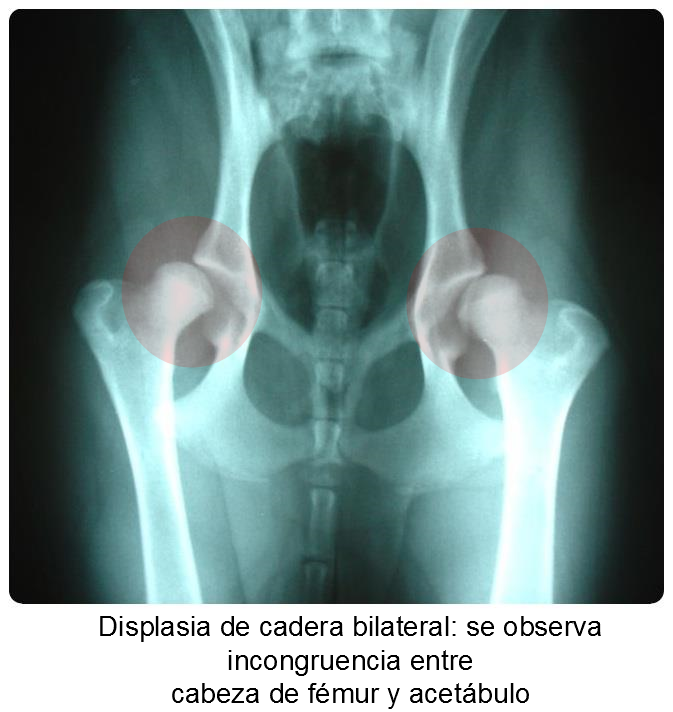
Hip dysplasia is a very common disease in large and giant breeds of dogs, which consists of defective development of this joint.

In it, the two bones that form the joint, femur and pelvis, do not adapt correctly due to different biomechanical imbalances produced during the growth of the animal. It is a hereditary disease, so the main solution to eradicate it is to avoid breeding these animals, although it is also important to control factors such as nutrition, weight or overexertion of the puppy during its growth as well as consanguinity in selective breeding.

The symptoms presented by the animals vary according to the severity of the dysplasia, from a slight lameness to the total inability of the animal to lead a normal life.

The diagnosis of this disease is not easy, since there is no method that allows it to be determined in all cases. The method accepted in Spain for certificate purposes is radiographic, although it has the disadvantage that it must be carried out when growth has finished, that is, after twelve months for most breeds.

Early diagnosis can prevent transmission

In the Doctoral Thesis of Beatriz Melo Alonso, defended at the University of León and directed by doctors José Manuel Gonzalo Orden and Mario Manuel Dinis, hip dysplasia has been investigated in one of our native breeds: the Burgos Pointer.
The result of this research has been worrying, since 59.3% of the animals studied suffer from hip dysplasia in its different degrees, with 18.6% severe dysplasia. This high percentage should alert the Burgos Perdiguero associations to try to eradicate it.

Early diagnostic

The drawback is that the aforementioned diagnostic technique is very late, and therefore, has a great emotional impact on owners.
For this reason, another part of the research has consisted of perfecting, for this breed, a new diagnostic technique developed in the United States called the PennHIP or distraction method, which consists of taking a specific X-ray and taking a measurement on it called the index of distraction.

This study has concluded that with the PennHIP method this disease can be predicted from four months of age, and throughout the animal’s growth, with the same reliability, in the Pointer, and it has even been possible to enunciate a formula with the which will know the degree of hip dysplasia that he will have in the future from the distraction index that he presents at four months. This system could reduce this disease, which has so much repercussion, both on the animal itself and on the owners.

Source: University of León

At Ortocanis we work to improve the lives of dogs with mobility problems, that includes dogs with some permanent physical disability as well as dogs that need rehabilitation. Marta Subirats, our collaborator, tells you about the rupture of the anterior cruciate ligament.

Of the four ligaments that make up the dog’s knee, rupture of the anterior cruciate ligament is one of the most common pathologies and the most frequent cause of secondary degenerative osteoarthritis in the knee joint. The functions of the cranial cruciate ligament are to limit internal rotation of the tibia and cranial displacement of the tibia relative to the femur and to prevent hyperextension of the knee.

protector-rodilla-perro (1)If your dog appears with an acute limp, does not want to put his paw on the ground, or seems to take a few steps and shrinks it, and his knee swells, it is possible that his anterior cruciate ligament is affected.

There is a predisposition in certain dogs to suffer this injury. On the one hand we find small-medium sized dogs (breed or not) with short legs and generally overweight and, on the other hand, large and giant breeds that, due to their morphology, have a tendency to suffer injuries to their ligaments. Among the latter we find the Labrador, Rottweiler, Neapolitan Mastiff, Boxer, etc. In any case, this is not a rule and any dog can have a ligament injury throughout his life.

Apart from the breeds mentioned, there are other factors such as being overweight, sedentary lifestyle, endocrine disorders, sports dogs that do not warm up properly, stairs, sudden climbs to the sofa or the car, or activities that subject the ligaments to micro-trauma and that finally end up damaging them with partial damage or total breakage.

There are two types of treatment, conservative and surgical, whether one or the other is chosen, the animal must carry out a correct recovery of that knee so that it becomes functional again and thus avoid recurrences.

The goals of recovery are to decrease pain, swelling, and lameness, to regain full mobility, muscle mass and strength, and control over the joint.

Physiotherapy varies depending on the animal and the type of treatment, conservative or surgical and, in the case of surgery, the type of intervention. There are surgeries that require more rest and stabilization than others. The recovery process is considered complete when the animal is able to perform daily activities and its knee is able to receive loads and movement without risk of re-injury.

It is important that your pet receives rehabilitation treatment by trained professionals who will choose the best techniques for a quick and effective recovery. Among the most used therapies to treat a crusader problem we find: massages, mobilizations, therapy with currents, ultrasounds, laser, aquatic therapy, balance and coordination training therapy.

During the recovery period it is important that:

– Take your pet on a leash during walks and avoid sudden departures towards other dogs and changes of pace, especially at the beginning of recovery. Your veterinarian or therapist will modify the intensity of the activity as the recovery period progresses.

– Keep the dog off slippery floors. A common cause is recurrence of the ligament tear accompanied by medial meniscus injury.

– Avoid ramps and stairs at the beginning of treatment in operated animals and in animals undergoing conservative treatment. Once rehabilitated, ramps can be used to help get on the sofa and in the car, since it is recommended that they do not do it alone, there could be a recurrence.

– It is recommended that they rest on soft and clean surfaces, but that they are firm enough to help the incorporation of the animal special mattress for dogs

– Keep the skin clean and dry.

– Correct diet and weight control. Being overweight harms the joints and generates moreprotector-rodilla-canina-perro articulada pain for the animal

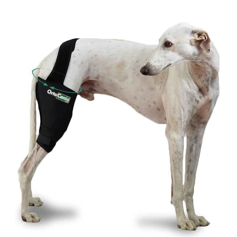
During recovery or in those animals in which knee instability may arise, the use of a hinged knee orthosis can benefit you and prevent recurrence or possible complications.

Knee orthoses can be used in cases where surgery is not possible or there is some impediment to perform it. These splints, designed exclusively for knee problems, allow progressively increasing degrees of flexion-extension while limiting unwanted movements, providing stability throughout the recovery.

Marta Subirats

Ortocanis physiotherapy technique

There are many treatments that exist for the rehabilitation of our little friends, one of the least known; thermotherapy!

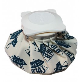
Thermotherapy is the application of heat to the body for therapeutic purposes. There are different ways of applying heat to the animal: heat packs , parafango, ultrasound , short wave, infrared , water vapor, contrast baths and moist heat.

It has therapeutic effects; It is anti-inflammatory, antispasmodic, analgesic, sedative, relaxing and decontracting.

At the cellular level, it increases metabolism, while at the level of the blood vessels, it acts by producing thermoregulation that, at the local level, will produce brief vasoconstriction followed by vasodilation, giving rise to hyperthermia. In addition to a vasomotor reaction.

At a deep level, hyperthermia also occurs. It will produce a reflex action as a consequence of the vasomotor modifications of the local application that will produce an improvement in cell nutrition, an anti-inflammatory and analgesic effect.bolsas-de-agua-caliente-o-hielo

On the heart it produces tachycardias and modifies blood pressure in local applications and if its application is increased, it produces an increase in the temperature of the stimulus and decreases blood pressure with an increase in volume.

In the blood, it produces an alkalinization of the blood pH, decreases coagulation, blood viscosity and, consequently, there is a greater lymphatic supply to the tissues.

On the skin there is an increase in temperature, with an increase in local circulation and decreased sensitivity.

The heat on the nervous system increases sensitivity in short-term applications, while if the duration is prolonged, it produces a decrease in sensitivity, sedation and analgesia.

At the muscular level, it produces relaxation, is antispasmodic, decreases excitability, increases tissue elasticity and decreases muscle tone. And, on the respiratory system, it produces an increase in the respiratory rate.

There are a number of circumstances in which heat cannot be applied:

  • Cardiac animals.
  • In acute inflammations 24-72 hours
  • Animals that present hypersensitivity or even allergic reactions
  • Areas:
    • Open, deep, or infected wounds
    • local infections
    • Burns
    • sensitivity disturbances
    • circulatory deficits
  • leishmaniasis

Heat can be applied once the acute phase of the injury or surgery has ended: fractures, contractures, tendinitis, dislocations, etc., or in chronic cases such as geriatric animals or those with osteoarthritis.

It is necessary to control the animal’s skin at all times and if the skin is very red or the animal is upset, the treatment should be stopped. It is necessary to place a towel between the animal and the heat, never place it directly, as there is a risk of causing skin burns.

There are special protectors or straps for a better fastening of the hot pack.

Orthocanis team

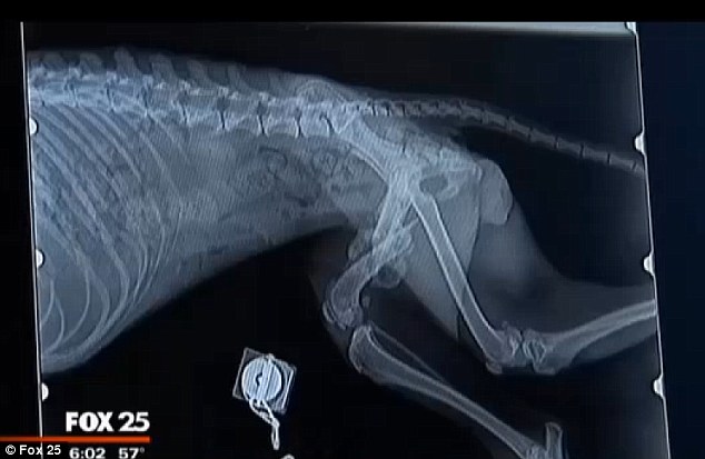
x-ray dog with fractures

A little more information from Ortocanis, do our articles help you?

Definition

Disc herniation is a neurological disease that affects the spine, when part of the intervertebral disc presses on or enters the spinal cord.

The dog has 7 cervical vertebrae, 13 thoracic, 7 lumbar, 3 sacral and, depending on the breed, 20-23 tail vertebrae. The intervertebral discs act as shock absorbers between the vertebrae that make up the spine and at the same time give it its mobility.

Two types of hernia and their symptoms

It mainly differentiates between two types of disc herniation: Protrusion and Discus Extrusion.

We speak of a Protrusion when the intervertebral disc moves and thus presses on the spinal cord but the spinal cord tissue is intact. The dog shows pain, walks awkwardly, sometimes with a hunched back and may drag its legs a little.

Extrusion means that the disc tissue has ruptured and disc material has entered and damaged the medulla. In this case the symptoms can be similar to that of the Protrusion but more serious. Depending on the location of the hernia, it causes paralysis of the forelimbs and/or hindlimbs. The dog loses sensitivity in those extremities, which manifests itself in the absence of pain, does not stand up and begins to crawl. It may also look like incontinence.

The most important thing in these cases is the rapid diagnosis and intervention of the veterinarian!!

A slight Protrusion can be treated with medication and almost absolute rest, however an Extrusion has to be operated to remove the disc material from the marrow.

But beware – each case of disc herniation is different and depends a lot on its location, to what degree it has pressed or damaged the spinal cord and how long it has been between the first symptom and diagnosis. That is why it is very important to choose a good neurologist.

Physiotherapy – recovery

In both cases, physiotherapy is very important for a good and faster recovery of the animal.

Various massage methods help stimulate the peripheral system and increase blood circulation. Passive joint movement prevents loss of mobility in affected limbs. Electrotherapy stops muscle atrophy and is the only passive way to increase muscle mass.

When the dog is already standing, several active exercises are applied to improve stability, balance and coordination.

The owner must take great care with the feeding of his dog during recovery and consult the veterinarian, since the loss of mobility and the same amount of food lead to weight gain – something that should be avoided in any case. Every extra gram makes it more difficult for the dog to get up and walk again.

In the event of a cervical hernia, the use of a harness is recommended to avoid sudden movements in this area and also to raise the food and water bowls so that the dog does not have to bend down too much.

andrea klein

animal physiotherapist

Collaborator of Ortocanis.com