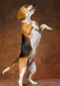
Hip dysplasia is a very common problem in certain breeds: BullDog, Bordeaux Doge, St. Bernard, Neapolitan Mastiff, German Shepherd, Rottweiler, Golden… all of them have an incidence above 20%.

Dysplasia is a multifactorial, multigenic and hereditary disease, that is, there are several factors that predispose and cause hip dysplasia, there are several genes involved in its appearance and it has a hereditary character.

Environmental factors are becoming more and more important in the development of hip dysplasia, the genetic factor is necessary to develop the disease but this is not the only factor. Genetics is a necessary but not exclusive factor, that is, you can have the genetic predisposition and not develop the disease but if you do not have a predisposition it is sure that it does not develop.

There are several degrees of dysplasia, and also those that appear when the dog is a puppy or those that give problem already in adulthood; but in this article we will focus on the treatment and specifically on the treatment of dysplasia to young dogs.

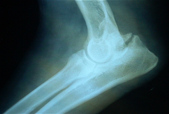
Classification of the degrees of dysplasia according to the OFA :

Grade I: minimal alteration with small subluxation and few degenerative changes.

Grade II: marked lateral subluxation of the femoral head, 25-50% of which is outside the acetabulum.

Grade III: 50-75% of the femoral head is outside the acetabulum; there are important degenerative changes.

Grade IV: dislocation of the femoral head with flattening of the acetabular border and femoral head; there are major degenerative changes.

The presentation in the young dog is puppy hip x-ray normally between 5 and 6 months and is marked by a significant limp.

A dog is not considered to be free of dysplasia until at two years of age already completed no problems or inconsistencies are observed in the control x-rays.

Food is one of the factors that predisposes to the appearance of hip dysplasia, Calcium-Phosphorus imbalances that must keep a correlation Ca1.6% – P1.1% and above all not overfeeding or providing excess proteins allows us to minimize the incidence of hip dysplasia. A hypocaloric diet from 3 months to 8 months protects dogs with rapid growth from dysplasia. Excess weight at 60 days is another factor that predisposes to the disease.

Hip dysplasia in the puppy usually debuts from 5 or 6 months, before it is not possible to observe any problem and the dog has been completely normal and has developed normally. The debut is usually presented as a sharp limp that prevents the dog from playing as it had done to date. We can observe changes in the desire to play, negative when going for a walk, to relate to other dogs or owners. Mood swings, frequent slips of the hind legs, discomfort and even refusal to be touched and the fact of “fleeing” from children in dogs that until a few days ago were playful and affectionate are frequent.

Sometimes when you reach 90% of the growth between 8 and 11 months the signs can be reduced and even disappear. Anyway the dysplasia remains and in many cases the problems reappear after a while and sooner rather than later signs of osteoarthritis appear in the hips.

The most common clinical signs are:

Lameness that may increase with exercise

Walking and jogging with hip swing

Morning stiffness

Difficulty getting up

Muscle atrophy

Refusal to move

Mood swings

Pain on palpation

Sign of Ortolani.

Although there are surgical methods: excision of the pectineus muscle, triple hip osteotomy, arthroplasty of the femoral head, osteotomy of the pubis, forage, hip prostheses most are practiced when the dog is young to supposedly decrease the possibility of secondary coxofemoral osteoarthritis in adulthood. The hip prosthesis should be reserved for severe cases and once the growth has finished.

Medical treatment is based on anti-inflammatories, we can start with natural anti-inflammatories, such as inflamex, which does not contain medicinal substances, if we do not obtain the expected results move to Aine’s and in extreme cases corticos are resorted to. We must include nutraceuticals especially chondroprotectors since they reduce the incidence of osteoarthritis and protect the articular cartilage. These are used in senior dogs in a very general way but are very useful as a joint protector in growing dogs, there are specific drug carriers for young dogs. Weight reduction, moderate and above all regular exercise are other basic points, as well as improvements in the environment and the fact of sleeping on a special mattress for older dogs and in a warm place away from humidity.

Canine physiotherapy can help a lot to better develop the muscles to reduce pain, to draw tensions and eliminate compensations that the dog has made with the wrong postures and antialgic positions. This will be based on TENS, ultrasound, therapeutic exercises, the use of hydrotherapy, laser, shock waves…

The main improvement in the environment is to sleep on a good therapeutic mattress, not to be cold or exposed to a lot of humidity, to use in winter a thermal coat for dogs, to be able to be a therapeutic canine blanket that can be used all year round.

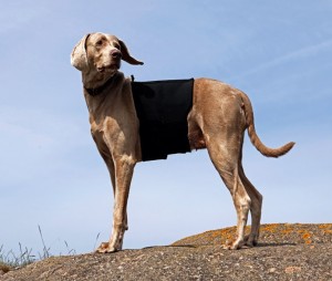
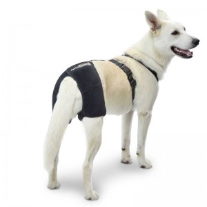
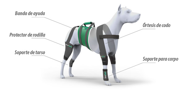
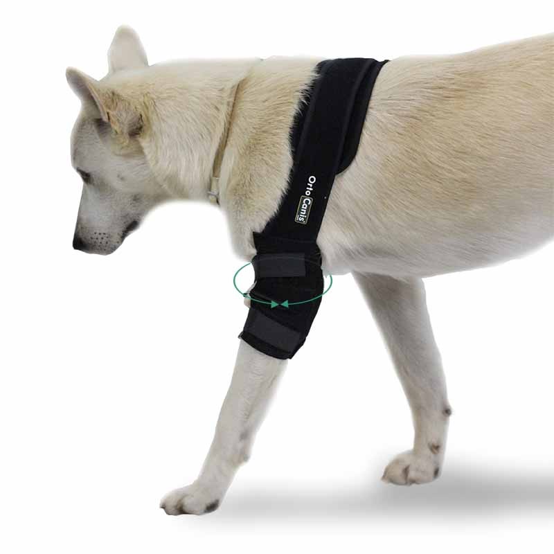
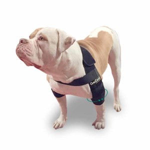
We can help our Dog with hip dysplasia, regular physical exercise can be very useful to improve muscle mass that better withstands poor joint congruence, avoid impacts, jumps or uncontrolled runs during the presentation of the picture are also important elements. Physiotherapy and massages allow you to always have the dog in a correct muscular state, and all the adjuvant treatments such as acupuncture, massages, reiki, bach flowers … they can also help with treatment. The latest novelty is the hip supports that help stabilize the pelvis, give support and greatly improve the quality of life of our pets.

Toni

Ortocanis.com Team

Deforming Spondylosis in Dogs

DEGENERATION OF THE SPINE IN DOGS

Deforming spondylosis is a degenerative, non-inflammatory disease of the spine, characterized by the production of bone spurs in the lower, sides, and upper area of the vertebrae of the spine. These spurs are simply caused by bone growths, usually growing in response to aging or injury.

In dogs, deforming spondylosis most often occurs along the spine, in the back of the chest, and in the upper vertebrae of the lower back. Older and large breed dogs are at higher risk of developing deforming spondylosis.

SYMPTOMS

*Patients are usually asymptomatic, bone growth can be felt by touching your pet before perceiving changes in its behavior in the wake of growth

  • Fracture of spurs or bridges can cause pain
  • Rigidity
  • Restricted movement
  • Pain

CAUSES

Repeated microtrauma – repeated pressure on the same joints or bones, through certain exercises or activities
Major traumas – the body responds when trying to grow new bone
*Predisposition to hered spurs

DIAGNOSIS

Your veterinarian will do a complete physical examination of your dog, including a biochemical profile, a complete blood count, a urinalysis, and an electrolyte panel, in order to rule out or confirm other diseases, such as cancer. You will need to give a complete history of your dog’s health, including background history of symptoms, onset of symptoms, and possible incidents that may have precipitated this condition.
X-ray images of the chest and abdomen (side view) are essential for the diagnosis of deforming spondylosis. X-rays reveal osteophytes (small bone growths) in the vertebrae, or in more advanced cases an osteophyte can be found as a bridge in the space between the vertebrae.

Your doctor may choose from several other types of tests in order to reach a definitive conclusion. A myelography uses the injection of a radiopaque substance to obtain an interior image; Computed tomography (CT) or magnetic resonance imaging (MRI) are also options. These procedures can help your veterinarian find a bone spur that may be putting pressure on your dog’s spinal cord or nerves (leading to neurological reactions).

TREATMENT

faja para el dolor de la espalda de perroUsually, patients with deforming spondylosis do not show abnormal external symptoms of initial bone growth. A neurological examination should be performed to rule out a pathology of the spine that requires surgery. Otherwise, if the growth has reached the point of damaging nerves and tissue, and your pet suffers intense pain, or if your veterinarian has decided on a surgical solution, your dog will be hospitalized. Under normal circumstances, when damage to the body is minimal, and your dog feels little discomfort and pain, the condition will be treated on an outpatient basis, with strict rest and prescription painkillers for home treatment. Pain medications are given after your meals. To speed up recovery and from four days after the intervention you can use thermal coats or spine girdles for dogs (consult your veterinarian). Acupuncture can also provide pain relief for some animals.

LIFE AND MANAGEMENT

Depending on the severity of the symptoms, your veterinarian will schedule checkups to track your dog’s progress. Only give pain medication when your dog is showing signs of discomfort (after a meal), and only in the exact amount prescribed, unless your veterinarian tells you otherwise. Drug or drug overdose is one of the most common causes of unintentional deaths in pets. You will need to provide a safe and quiet place for your dog to rest, away from other pets and active children. During this time, limit yourself to slow walks around the neighborhood. When your dog shows no signs of discomfort for several weeks he can slowly return to normal activity.

Source: Venfido
www.ortocanis.com

The phenotypic evaluation of hips done by the Orthopedic Foundation for Animals falls into seven different categories. Those categories are Normal (Excellent, Good, Fair), and Dysplastic (Mild, Moderate, Severe). Once each of the radiologists classifies the hip into one of the 7 phenotypes above, the final hip grade is decided by a consensus of the 3 independent outside evaluations. Examples would be:

  1. Two radiologists reported Excellent, one Good—the final grade would be Excellent
  2. One radiologist reported Excellent, one Good, one Fair—the final grade would be Good
  3. One radiologist reported Fair, two radiologists reported Mild—the final grade would be Mild

The hip grades of Excellent, Good and Fair are within normal limits and are given OFA numbers. This information is accepted by AKC on dogs with permanent identification (tattoo, microchip) and is in the public domain. Radiographs of Borderline, Mild, Moderate and Severely dysplastic hip grades are reviewed by the OFA radiologist and a radiographic report is generated documenting the abnormal radiographic findings. Unless the owner has chosen the open database, dysplastic hip grades are not in the public domain.

Excellent

Excellent: this classification is assigned for superior conformation in comparison to other animals of the same age and breed. There is a deep seated ball (femoral head) which fits tightly into a well-formed socket (acetabulum) with minimal joint space. There is almost complete coverage of the socket over the ball.

Excellent Hips

Good

Good: slightly less than superior but a well-formed congruent hip joint is visualized. The ball fits well into the socket and good coverage is present.

Good Hips

Fair

Fair: Assigned where minor irregularities in the hip joint exist. The hip joint is wider than a good hip phenotype. This is due to the ball slightly slipping out of the socket causing a minor degree of joint incongruency. There may also be slight inward deviation of the weight-bearing surface of the socket (dorsal acetabular rim) causing the socket to appear slightly shallow. This can be a normal finding in some breeds however, such as the Chinese Shar Pei, Chow Chow, and Poodle.

Fair Hips

Borderline

Borderline: there is no clear cut consensus between the radiologists to place the hip into a given category of normal or dysplastic. There is usually more incongruency present than what occurs in the minor amount found in a fair but there are no arthritic changes present that definitively diagnose the hip joint being dysplastic. There also may be a bony projection present on any of the areas of the hip anatomy illustrated above that can not accurately be assessed as being an abnormal arthritic change or as a normal anatomic variant for that individual dog. To increase the accuracy of a correct diagnosis, it is recommended to repeat the radiographs at a later date (usually 6 months). This allows the radiologist to compare the initial film with the most recent film over a given time period and assess for progressive arthritic changes that would be expected if the dog was truly dysplastic. Most dogs with this grade (over 50%) show no change in hip conformation over time and receive a normal hip rating; usually a fair hip phenotype.

Mild

Mild Hip Dysplasia: there is significant subluxation present where the ball is partially out of the socket causing an incongruent increased joint space. The socket is usually shallow only partially covering the ball. There are usually no arthritic changes present with this classification and if the dog is young (24 to 30 months of age), there is an option to resubmit an radiograph when the dog is older so it can be reevaluated a second time. Most dogs will remain dysplastic showing progression of the disease with early arthritic changes. Since HD is a chronic, progressive disease, the older the dog, the more accurate the diagnosis of HD (or lack of HD).

Mild Dysplasia

Moderate

Moderate Hip Dysplasia: there is significant subluxation present where the ball is barely seated into a shallow socket causing joint incongruency. There are secondary arthritic bone changes usually along the femoral neck and head (termed remodeling), acetabular rim changes (termed osteophytes or bone spurs) and various degrees of trabecular bone pattern changes called sclerosis. Once arthritis is reported, there is only continued progression of arthritis over time.

Moderate Dysplasia

Severe

Severe Hip Dysplasia: assigned where radiographic evidence of marked dysplasia exists. There is significant subluxation present where the ball is partly or completely out of a shallow socket. Like moderate HD, there are also large amounts of secondary arthritic bone changes along the femoral neck and head, acetabular rim changes and large amounts of abnormal bone pattern changes.

Other Hip Dysplasia Registries—An Approximation

OFA FCI (European) BVA (UK/Australia) SV (Germany)
Excellent A-1 0-4 (no > 3/hip) Normal
Good A-2 5-10 (no > 6/hip) Normal
Fair B-1 11-18 Normal
Borderline B-2 19-25 Fast Normal
Mild C 26-35 Noch Zugelassen
Moderate D 36-50 Mittlere
Severe And 51-106 Schwere

Font: Orthopedic Foundation For Animals

Although genetics is the determining part of dysplasia by 99% percent, it is nutrition that is the most important part. Once you have diagnosed dysplasia you can NOT do anything genetically, you only have to influence nutrition and physiotherapy. It’s that simple.

I teach you five key secrets in nutrition to make your best amig@’s dysplasia more bearable. In addition to an Expert in Canine Nutrition, do not forget to also consult one in canine physiotherapy, it can help you a lot.

Nutrición Ortocanis

GRAINS IN THE DIET

Unfortunately the vast majority of commercial veterinary diets for dogs are bad, why? Because they have a lot of carbohydrates in the form of grains and / or cereals, they do it to reduce costs, they are cheaper than animal protein, the nutrient that your dog really needs.

Dietary grains or cereals have been shown in dogs to promote continuous and exaggerated insulin secretion* as well as joint inflammation. I remind you that your dog does not have the physiology to split starches, carbohydrates, that is, grains and cereals. It is a carnivore, do not forget, do not feed it as if it were cow or chicken.

GLYCOSAMINOGLYCANS

Don’t panic with the name. They are nutrients that promote cartilage health. Patients with dysplasia problems (and arthritis in general) have shown that they can absorb toxic compounds or substances, some of which affect the joints.

How can we lessen this? If you give your perr@ some cartilage (rich in glycosaminoglycans) in the diet, these are poorly absorbed and remain in the intestinal lumen.

Glycosaminoglycans are complex carbohydrates that have the ability to adhere some of these toxic substances on their surface while they are in the lumen of the intestine, and thus be excreted in the excrement without passing into the bloodstream and therefore prevent their arrival and implantation in the joints.

A substitute for cartilage? There are nutritional supplements based on glycosaminoglycans, in short, they are more practical and come concentrated in a pill. There are many brands on the market: Cosequin, Synoquin, Hyal oral…

BELOW OR ABOVE WEIGHT?

If I have to choose to be slightly above my weight, I prefer the latter, and the same applies to my dog.

As an Expert in Canine Nutrition, this topic, that of weight, is one of the most recurrent. Owners of puppies of large and/or molossian breeds (mastiffs, Rottweiler, Siberians, shepherds, doges…) want “stuffed” puppies growing as much as possible. Gross error.

Remember, if you want a healthy dog orthopedically weight is key (it also applies to us) Have you seen overweight wolves, lions or hyenas in the wild?

How do you know if your dog is at an appropriate weight? The best allies for this are sight and touch. I recommend you visit the following link so that you learn how to determine the body condition of your dog.

BONES IN THE DIET

There are many myths in dog nutrition, created, above all, by the pet food industry. I remind you that your dog is a carnivore, every carnivore eats bones in nature, moose, rabbits, all kinds of birds (such as chicken or chicken) and ALL BONES, absolutely all, splinter and do not die!

It refreshes your memory. Commercial foods (pellets) have been massively and widespread in the last two decades. Before everyone gave homemade food (leftovers) including bones.

Bone is a mine of vitamins, fats and minerals of the highest quality, do not forget that bone is mainly made up of minerals such as calcium, phosphorus, fluoride, magnesium … water, and organic matter such as collagen. All of these nutrients are key to bones and joints, among other organs.

By the above I do not mean that you start introducing bones into the diet without knowing. COOKED BONES are the dangerous ones. The best diets are homemade, and if they include bones (RAW) occasionally, better.

NUTRITIONAL SUPPLEMENTS

If I am given the choice between providing a quality diet for a dog without supplements (the best diets are well-made homemade ones) or a poor quality diet and a quality supplement, I stay with the first without a doubt.

Now, if I can choose for my dog a quality homemade diet, alternated with a commercial diet, also of quality, and added with a specific supplement for his dysplasia problems Bingo!

With the arrival of orthomolecular nutrition (specific nutrients for specific situations) and the advancement of science, many nutrients appear that at the molecular level have a positive impact on the patient with dysplasia problems.

From vitamin E, bioflavonoids to omega three and enzymes. There are more and more of these nutrients for multiple situations. Regarding the subject we are dealing with, dysplasia, it would be something that you should not overlook and ask for advice from a specialist in the subject.

Carlos Alberto Gutierrez / Veterinarian collaborating with Ortocanis.com

Treatment of hygroma of the elbow

Elbow hygroma is a disease that mainly affects large short-haired dogs, such as Big Danes, Greyhounds and Dalmatians. In dogs with elbow hygroma, a fluid-filled sac appears on one or both of the dogs’ elbows. In rarer cases, hygromas can develop in the hock. In many cases, elbow hygromas do not cause pain or difficulty. However, they can become infected, which can be painful for the dog and requires treatment. Other animals susceptible to developing hygromas are dogs that are not very active or are recovering from illness or injury.

Causes
Hygromas develop because of recurrent trauma to a dog’s elbow or elbow. For example, resting constantly on wood, cement, or other hard surfaces can cause stress on the joint and cause hygroma. This condition mainly affects large dogs, as there is a greater weight on their elbow joints when they are lying down.Hygroma codo

Bandage
In some cases, bandaging a dog’s elbows can help prevent hygromas from getting worse. Bandages will prevent contact with hard surfaces and in turn avoid problems caused by hygroma. In addition, some products such as the canine elbow protector or the dog knee protector will prevent the hygroma from having additional contact with the hard ground. Ask your veterinarian how you can help your dog.

Drainage
Some veterinarians recommend that hygroma fluid be drained with a needle or syringe. Getting the fluid out is not considered an effective treatment because the needle can cause an infection. In addition, drainage can only improve the condition temporarily. If the dog continues to lie on hard surfaces, the hygroma will become larger, that is, the drainage will have to be done on a regular basis.

Surgery
Some veterinarians may recommend surgery to treat elbow hygroma, especially in cases where there is infection or when the hygroma has ulcerated. In surgery, the skin must be drained and removed. Since hygromas can grow quite large, it may be necessary to make skin grafts to cover the affected area. Healing from surgery takes about a month, and the dog will need to wear a splint during recovery to protect the affected area.

Prevention
The best way to prevent your dog from developing an elbow hygroma, or to prevent an existing one from worsening, is to provide the dog with a soft surface for him to rest and sleep. If there is no carpeted area available, make the dog a soft bed and leave it where he likes to lie down. In the event that the dog is inactive because he is recovering from an illness or injury, have him get up and move (at least enough to change the position he is lying in) several times a day.

Written by Anna Aronson

source: eHow

Elbow dysplasia is a very common degenerative disease in young dogs. The elbow of dogs is one of the most congruent and stable joints of the body, allowing, due to its complexity, two axes or degrees of supination-pronation movement of the forearm and flexion-extension. Its complexity is given by its composition: humeroradial joint, humeroulnar and, proximal radioulnar.

Elbow dysplasia was initially used to describe the non-union of the anconeal process (AUP). Currently, osteochondritis dissecans (OCD) of the medial condyle of the humerus, the fragment of the coronoid process (FPC) and, the incongruence of the elbow (INC) are also included within this term. When one of these ossification defects occurs in an elbow, inflammation originates and over time an osteoarthritis is triggered in which cartilage degeneration occurs; for that reason, all these conditions are commonly associated with osteoathrosis of this joint and are an important cause of pain and claudication of the forelimbs in large and giant breed dogs such as the German Shepherd, Labrador, St. Bernard, Rottweiler, Neapolitan Mastiff, among others.

Of genetic origin multifactorial, especially in OCD and FPC. It affects males more than females and can occur uni- or bilaterally. The genetic component is the one that has the greatest influence although, the appearance of this pathology can also occur due to food, weight, environment, quality of ligaments, a lot of physical exercise or trauma.

The first symptoms may occur at 4-5 months when the dog shows exercise intolerance, lameness when starting a movement or after prolonged exercise. There are dogs that do not show signs of affection in the elbow until advanced ages where the process of osteoarthritis is very evolved. Others manage to maintain a normal degree of activity throughout their lives.

The fact of making a premature radiological diagnosis makes it possible to establish an adequate treatment and avoids the formation of osteoarthritis that produces pain and functional limitation of the elbow throughout the life of the animal. The diagnosis can be complemented with diagnostic tests such as CT or MRI

The evolution depends on the degree and type of injury, but it is usually unfavorable without surgery. Surgical treatment is good if degenerative changes in the joint have not yet occurred. In any case it is necessary to perform a good rehabilitation in order to:

  • Speed up the recovery process
  • Eliminate pain and inflammation
  • Decrease lameness
  • Maintain and/or improve range of motion
  • Maintain muscle tone, mass and strength
  • Minimize or slow down the effects of joint degeneration – osteoarthritis
  • Avoid compensation at the level of the neck, spine and extremities
  • Give the maximum capacities so that the animal is functional and that it, with a good quality of life

Physiotherapy treatment varies depending on the animal and the state of the lesion. It is important to start as soon as possible with the treatment so that it is effective and, to avoid drying them as reduced mobility and / or chronic pain.

The animal goes through different phases until its full recovery. It is essential to gradually achieve the objectives set. The recovery process is terminated when the animal is able to perform daily activities.

During the first three days after the intervention, it is important to act on inflammation and pain and prevent muscle atrophy and decrease in the joint arch from appearing. For this, passive techniques are used that reduce inflammation, produce analgesia and help maintain tone, mass and the arc of mobility. Among these techniques there are electrotherapy (segmental TENS and muscle electrostimulation), massage, passive mobilizations and cryotherapy (cold).

In older dogs or dogs that have not been intervened, the objectives will be the same as in animals that have gone through an intervention. It is important to eliminate pain because, with pain you can not work.

It is important from the beginning to massage and move the affected elbow as long as there is no veterinary contraindication and, respecting in the case of fixation, the period of healing and union of the fixed parts. Massaging and moving the affected area and limb helps maintain mobility, prevents loss of mass and tone and works the proprioceptors.


A gentle mobilization combined with different massage techniques help decrease inflammation and reduce pain.

With TENSat the segmental level we can produce analgesia and decrease the amount of drugs administered. There are animals that have intolerance to certain drugs that produce analgesia and with TENS the pain can be reduced. TenS can also be used directly on the injured or operated area, as long as there is no osteosynthesis material underneath, since an internal burn could occur.

Muscle electrostimulationhelps prevent the onset of atrophy and maintain muscle mass and tone. With electrical stimuli we can stimulate nerve conduction.

At the beginning and end of the session the coldis used since it has properties that act on the decrease of the inflammatory response, edema and pain.

From the fourth day and during the next two weeks when the inflammation and pain have disappeared it is time to introduce simple active exercises such as shaking hands or small walks on a leash to force the animal to make an equal support with the four limbs and thus, prevent a decompensation between limbs from appearing due to not having a correct support on the ground. The walks is an exercise that increases the duration until full recovery.

Once the stitches have been removed, the animal can be introduced into the water. The advantages of water are used to improve recovery. Hydrotherapy (underwater treadmill) facilitates the station of the animal without loss of balance and, thanks to flotation, without having to support all its weight. In addition, flotation allows animals with bone pain and low muscle mass to work. The pressure of the water exerted on the body of the animal increases the sensitivity and decreases inflammations and edemas. The work in the water, underwater tapes or swimming increases as the animal recovers. In addition, with water, we can recover the motor pattern, increase mass, tone and strength, work on respiratory capacity and maintain and / or improve mobility.

Once the acute phase has passed 48-72 hours and without risk of infection or inflammation, heat can be introduced that helps to elastify the tissues, decreases pain and increases vascularization among others.

The use of boards, plates, balls and trampolines are important to work on balance, proprioception and above all the integration of the affected limb.

It is already in the last phase, from two weeks, when the dog has integrated the gait pattern, exercises are performed to improve the quality of movement. They are more complex active exercises to integrate the affected limb or limbs. With active and proprioception exercises it is possible to increase muscle tone, mass and strength; coordination and balance and range of motion are worked on. Rails with different surfaces, cones, bars, circuits, up and down stairs and ramps (staircase with inclined plane) are used.

Throughout the recovery treatment and in animals with developed osteoarthritis it is essential to reduce the weight on the joints of the elbows. For this purpose , special support harnesses for elbows are used. In addition to decreasing weight, pain is reduced and does not hinder movement, the animal feels more comfortable; the joint is protected at all times from chafing and blows and helps maintain the heat that the animal gives off, which leads to a relief of the affected area.

At home, special care should be taken for animals suffering from elbow conditions. This care is necessary during and after treatment:

  • Avoid slippery floors
  • Avoid ramps and stairs at the beginning of treatment in operated animals and in animals that do conservative treatment. Once rehabilitated, ramps can be used to help get on the sofa and the car, since it is recommended that they do not do it alone, there could be a recidivism.
  • It is recommended that they rest on soft and clean surfaces, but that they are firm enough to help the incorporation of the animal thermal mattress for dogs
  • Keep skin clean and dry skin
  • Use special plates at your height so as not to strain the elbow joints
  • Correct diet and weight control. Being overweight harms the joints and generates more pain for the animal

It is very important to create an exercise routine and environment to help keep the animal comfortable and with quality of life.

Orthocanis Team

Electrostimulation for muscle potentiation

Electrostimulation is a very useful tool to enhance the muscles of a dog that has suffered an injury or surgical intervention and as a result has muscle atrophy.

We can use electric current to stimulate the muscles, enhance it and make it work, it is specifically indicated in cases of muscle atrophy, and especially in cases where the dog can not do active work. If the dog can do active work, electrotherapy will be a support, never the main source of work.

Form of the impulse that we will use will be the Rectangular, symmetrical biphasic the main authors indicate it: Thepaut Mathieu 1992, Kramer 1984, Bircan 2002

pack-tens-para-perros

Human electrostimulation apartments can be adapted to electrostimulate dogs without too many problems, but we must ensure that we can vary the frequencies, and especially the amplitudes of the electrical impulse to adapt well to the characteristics of the dog.
There are no good studies of Cronaxia in dogs, the approximate values are 0.3 human, 0.2 Horses and in dogs by studies of less depth intuit similar values;
Sawaya – Meallier 2006, Brodart 1998, Coarasa 1999, Ramon 2007
The Intensity according to Hultaman 1983, and Ogino 2002 should be elevated with visible contraction, without reaching the limit of pain but discomfort. The dog must comfortably withstand the session but the muscles must work intensely.

The frequency will be marked according to the objective, various authors validate this option: Pougheon 1992, Busko 1989, Vanderthommen 2002.

Objective Frequency Treatment time Standby time
Relaxation: 5 Hz Continuous 0
Heating: 5 Hz Continuous 0
“Endurance”: 10-20 Hz 9 2
Atrophy: 33 Hz 6 6
Strength: 50- 100 Hz 5 25
Explosive force: 100-200 Hz 3 30

The parameters may vary slightly according to individual cases.
Rest time can be adjusted if it is active, it is advisable to use ramps before strong muscle contraction.
The intensity should always be the maximum with relative comfort.
Total time between 10 and 30 minutes depending on the phase of the injury, fatigue the muscles excessively does not help to enhance the muscles correctly.

It is important to have a device that meets all the requirements, if it is going to be used by a canine rehabilitation center or veterinary hospital, a more professional desktop apartment is recommended and with it we can use specific programs for each of our patients.

The use of rubber electrodes and contact gel is equally recommended in animals with hair.


Orthocanis Veterinary Team

Baner silla 728x90

It is one of the most used instruments in physiotherapy. It is a device that emits acoustic waves of much higher frequencies than those audible by humans. The frequency used as a therapeutic tool is 1×10⁶ Hertz, i.e. 1 Mega-Hertz (MHz), so they are not audible by any mammal.
Usually in veterinary clinics and hospitals, ultrasound is used for ultrasound scans that use the same type of wave. The difference is power, frequency and application time.
In therapeutics we use frequencies of 1MHz for deep treatments, up to 8 cm and frequencies of 3MHz for more superficial problems. The power ranges between 0.2 and 3 Watts / square centimeter.

Effects on tissues:

The main effect of ultrasound on tissues is anti-inflammatory. We usually use it in tendons, joints or inflamed muscles; it has excellent results in both acute injuries and chronic injuries, although we must adjust the powers.

The analgesic effect is another of the most sought after in rehabilitation, normally when we deflate a structure we manage to reduce the pressure in the nociceptors that are the receptors in the body that send the painful signals, by reducing the pressure in these receptors, we reduce their stimulation and therefore decrease the intensity of the signals they send until they disappear. If there is no sign of pain, there is no perception of it.

soporte para perro con displasia de caderaWhen we have a fibrosis in the different soft tissues: muscles, tendons or ligaments, we can apply continuous and then pulsating ultrasound at maximum power. This way we will find a good defibrosing effect.

Another of the classic applications of ultrasound is the application in muscle contractures, with ultrasound we can reduce and even eliminate them.

Continuous ultrasound generates heat by the vibration of the molecules and both the pulsating and the continuous increase the permeability of the membrane, which is what favors together with the mobilization of the molecules the anti-inflammatory effect.

Application:

Ultrasound must be applied by moving the head all the time that the treatment lasts, either making small circles or following the direction of the treated tissues rectilinearly. If we do not do so, especially in continuous mode, we can damage tissues and produce significant burns.
It is necessary to use a means of contact, either contact gel or directly in direct immersion (sub-aquatic ultrasound).
It is also important to take into account the hair of the animal, this hinders the transmission of ultrasound and therefore it is very interesting to be able to shave the dog before applying the ultrasound sessionperro-multimedia-600x300_6

Approximate time between 5 and 15 minutes
Power between 0.2 and 3 Wats/cm2
There are different head measurements depending on the needs.

Directions:

Ultrasounds can be used in any pathology of the dog that occurs with joint or soft tissue pain, such as tendonitis, bursitis, arthritis, bruises or major bruises.

We can also use ultrasound in chronic problems such as hip dysplasia, elbow dysplasia, knee osteoarthritis, or hip osteoarthritis.

All surgical interventions produce an inflammation of the tissues that have been operated, ultrasound is a very good tool to control inflammation and post-surgical problems such as rupture of the cranial cruciate ligament, patella dislocation or others.

Ortocanis technical team

Baner silla 728x90


Hip dysplasia Irresponsible breeders, feeding, environment?

Currently, with only three months of age it is possible to know the existence of small anomalies in the conformation of the hip/femur joint, which will lead to dysplasia.

Origin of hip dysplasia in dogs:

In colloquial language (today we avoid veterinary terms) hip dysplasia is a “failure” in the head joint of the femur-hip. If the head of the femur is not lodged in the hip perfectly, there is a deterioration of the cartilage that protects the joint, and that deterioration is degenerative and irreversible. But why does hip dysplasia occur?
Genetic inheritance. Hip dysplasia is inherited, and if breeders do not perform the necessary tests to know that their dogs are free (certified X-ray) and that previous generations too, puppies can suffer from it. Many breeders (and more particular) ignore these x-rays (eye, there is no breed safe from the disease). Hopefully they include a clause in the sales contract in which they will give you a puppy if you prove that the one you have bought has dysplasia (as if they were appliances).
Environmental factors. In the period of growth (until the year, but especially critical the first six months of life), slippery floors, sudden exercises, jumps … Puppies with a limit hip can aggravate their situation if care is not taken in these critical months, and vice versa, they will be able to lead a perfectly normal life if they develop correctly in these months (even if their hips are not perfect).
Feeding. The months in which dysplasia develops are those of growth, and the slower the puppy grows the better. Foods very high in protein have been linked to the onset of dysplasia. Chondroprodectors help during growth (in predisposed individuals or breeds, always under veterinary supervision).
About prevention in line with the above, if the breeder is responsible and has all the controls done we still can not sing victory. It is very important that the puppy has a good diet according to his growth needs, which does not get fat (the image we all have of a rolly puppy is typical, but not healthy), supported by chondroprotectors if necessary, that the exercise is restrained (avoiding strange movements, and especially jumps and forced postures of the back three), be careful with the floors of the house (if they are slippery it is not a bad idea to get some old carpets that last us a few months).

Some exercises and “tricks” are very demanding with the hip, and therefore dangerous in puppies and young dogs.

And the greatest prevention: radiography There are many puppies that can limp for causes that have nothing to do with dysplasia, and in the same way, there are asymptomatic with serious femur and hip problems. The plate is painless, economical, and the only truly reliable method. At present we can know the state of the hips of our puppy from as early as three months (PennHip method), so that conservative treatments can be established, or in the case of an intervention being necessary, which is not drastic but reconstructive, preserving the joint. Until the year of age it is not possible to ensure that the hip has had a perfect development and, therefore, it will not be until then when the dog can start in canine sports (“start” is to go little by little) performing more demanding exercises with his body.

Source: www.doogweb.es

www.ortocanis.com


Elbow dysplasia in dogs

Elbow dysplasia is a very common degenerative disease in young dogs.

The elbow of dogs is one of the most congruent and stable joints of the body, allowing, due to its complexity, two axes of or degrees of supination-pronation movement of the forearm and flexion-extension. Its complexity is given by its composition: humeroradial joint, humeroulnar and, proximal radioulnar.

Elbow dysplasia was initially used to describe the non-union of the anconeal process (AUP). Currently, osteochondritis dissecans (OCD) of the medial condyle of the humerus, the fragment of the coronoid process (FPC) and, the incongruence of the elbow (INC) are also included within this term. When one of these ossification defects occurs in an elbow, inflammation originates and over time an osteoarthritis is triggered in which cartilage degeneration occurs; for that reason, all these conditions are commonly associated with osteoathrosis of this joint and are an important cause of pain and claudication of the forelimbs in large and giant breed dogs such as the German Shepherd, Labrador, St. Bernard, Rottweiler, Neapolitan Mastiff, among others.

protector-codo-canino

Of multifactorial genetic origin, especially in OCD and FPC. It affects males more than females and can occur uni- or bilaterally. The genetic component is the one that has the greatest influence although, the appearance of this pathology can also occur due to food, weight, environment, quality of ligaments, a lot of physical exercise or trauma.

The first symptoms may occur at 4-5 months when the dog shows exercise intolerance, lameness when starting a movement or after prolonged exercise. There are dogs that do not show signs of affection in the elbow until advanced ages where the process of osteoarthritis is very evolved. Others manage to maintain a normal degree of activity throughout their lives.

The fact of making a premature radiological diagnosis makes it possible to establish an adequate treatment and avoids the formation of osteoarthritis that produces pain and functional limitation of the elbow throughout the life of the animal. The diagnosis can be complemented with diagnostic tests such as CT or MRI

The evolution depends on the degree and type of injury, but it is usually unfavorable without surgery. Surgical treatment is good if degenerative changes in the joint have not yet occurred. In any case it is necessary to perform a good rehabilitation in order to:

  • Speed up the recovery process
  • Eliminate pain and inflammation
  • Decrease lameness
  • Maintain and/or improve range of motion
  • Maintain muscle tone, mass and strength
  • Minimize or slow down the effects of joint degeneration – osteoarthritis
  • Avoid compensation at the level of the neck, spine and extremities
  • Give the maximum capacities so that the animal is functional and that it, with a good quality of life

Physiotherapy treatment varies depending on the animal and the state of the lesion. It is important to start as soon as possible with the treatment so that it is effective and, to avoid drying them as reduced mobility and / or chronic pain.

The animal goes through different phases until its full recovery. It is essential to gradually achieve the objectives set. The recovery process is terminated when the animal is able to perform daily activities.

During the first three days after the intervention, it is important to act on inflammation and pain and prevent muscle atrophy and decrease in the joint arch from appearing. For this, passive techniques are used that reduce inflammation, produce analgesia and help maintain tone, mass and the arc of mobility. Among these techniques there are electrotherapy (segmental TENS and muscle electrostimulation), massage, passive mobilizations and cryotherapy (cold).

In older dogs or dogs that have not been intervened, the objectives will be the same as in animals that have gone through an intervention. It is important to eliminate pain because, with pain you can not work.

It is important from the beginning to massage and move the affected elbow as long as there is no veterinary contraindication and, respecting in the case of fixation, the period of healing and union of the fixed parts. Massaging and moving the affected area and limb helps maintain mobility, prevents loss of mass and tone and works the proprioceptors.

ortesis-codoA gentle mobilization combined with different massage techniques help decrease inflammation and reduce pain.

With TENSat the segmental level we can produce analgesia and decrease the amount of drugs administered. There are animals that have intolerance to certain drugs that produce analgesia and with TENS the pain can be reduced. TenS can also be used directly on the injured or operated area, as long as there is no osteosynthesis material underneath, since an internal burn could occur.

Muscle electrostimulationhelps prevent the onset of atrophy and maintain muscle mass and tone. With electrical stimuli we can stimulate nerve conduction.

At the beginning and end of the session the coldis used since it has properties that act on the decrease of the inflammatory response, edema and pain.

From the fourth day and during the next two weeks when the inflammation and pain have disappeared it is time to introduce simple active exercises such as shaking hands or small walks on a leash to force the animal to make an equal support with the four limbs and thus, prevent a decompensation between limbs from appearing due to not having a correct support on the ground. The walks is an exercise that increases the duration until full recovery.

Once the stitches have been removed, the animal can be introduced into the water. The advantages of water are used to improve recovery. Hydrotherapy (underwater treadmill) facilitates the station of the animal without loss of balance and, thanks to flotation, without having to support all its weight. In addition, flotation allows animals with bone pain and low muscle mass to work. The pressure of the water exerted on the body of the animal increases the sensitivity and decreases inflammations and edemas. The work in the water, underwater tapes or swimming increasesas the animal recovers. In addition, with water, we can recover the motor pattern, increase mass, tone and strength, work on respiratory capacity and maintain and / or improve mobility.

Once the acute phase has passed 48-72 hours and without risk of infection or inflammation, heat can be introduced that helps to elastify the tissues, decreases pain and increases vascularization among others.

The use of boards, plates, balls and trampolines are important to work on balance, proprioception and above all the integration of the affected limb.

It is already in the last phase, from two weeks, when the dog has integrated the gait pattern, exercises are performed to improve the quality of movement. They are more complex active exercises to integrate the affected limb or limbs. With active and proprioception exercises it is possible to increase muscle tone, mass and strength; coordination and balance and range of motion are worked on. Rails with different surfaces, cones, bars, circuits, up and down stairs and ramps (staircase with inclined plane) are used.

Throughout the recovery treatment and in animals with developed osteoarthritis it is essential to reduce the weight on the joints of the elbows. For this purpose , special support harnesses for elbows are used. In addition to decreasing weight, pain is reduced and does not hinder movement, the animal feels more comfortable; the joint is protected at all times from chafing and blows and helps maintain the heat that the animal gives off, which leads to a relief of the affected area.

At home, special care should be taken for animals suffering from elbow conditions. This care is necessary during and after treatment:

  • Avoid slippery floors
  • Avoid ramps and stairs at the beginning of treatment in operated animals and in animals that do conservative treatment. Once rehabilitated, ramps can be used to help get on the sofa and the car, since it is recommended that they do not do it alone, there could be a recidivism.
  • It is recommended that they rest on soft and clean surfaces, but that they are firm enough to help the incorporation of the animal special mattress for dogs
  • Keep skin clean and dry skin
  • Use special plates at your height so as not to strain the elbow joints
  • Correct diet and weight control. Being overweight harms the joints and generates more pain for the animal

It is very important to create an exercise routine and environment to help keep the animal comfortable and with quality of life.


You can also check:

Can chondroprotectors help?

Elbow dysplasia in dogs

Hip dysplasia in dogs

Baner silla 728x90