What does magnetic resonance imaging involve?
Magnetic resonance imaging is a powerful diagnostic tool that involves the use of magnetic fields. It is used to obtain non-invasive digitalised anatomical images without harmful effects, as it does not use ionising radiation, which is necessary to perform X-Rays and Computerised Axial Tomography scans (CAT scans).
It also has the ability to produce images in any plane without having to reposition the animal. It also provides better spatial resolution, a greater range of tissue contrast and increased sensitivity to pathological processes.
Nuclear magnetic resonance imaging, or MRI, is a safe diagnostic examination that provides an image of the inside of the body that is clearer than the ones offered by many other diagnostic examinations. Magnetic resonance produces two or three dimension images. It does not use X rays. A contrast agent can also be used to help to see the images better.
What are the advantages of resonance?
Magnetic resonance imaging helps to detect illnesses and treat them early on. It provides detailed information quickly and can reduce the need for certain types of diagnostic surgery.
In general, the vet will recommend an MRI scan if he/she suspects the presence of intracranial injuries or spinal cord injuries, but the scope of the diagnosis provided by the MRI scan is much wider.
In which cases are MRI scans recommended?
Below is a list of clinical settings where the use of resonance is recommended:
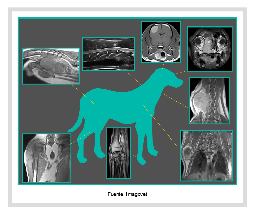
THE BRAIN, THE CEREBELLUM AND THE BRAIN STEM
When there are suspected brain injuries such as seizures, cranial nerve deficits, vestibular disease, nystagmus, ataxia, central blindness, circling, changes in behaviour and personality, altered mental status, locomotor problems, posture problems, eating disorders, diabetes insipidus, thermoregulatory deficit, changes in sleeping patterns (usually this involves sleeping too much) cardiovascular disorders (vasodilation is common), respiratory disorders, polyphagia, polydipsia, tremors, hyperkinesia.
SPINAL CORD
In animals with clinical signs like acute or chronic paresis/plegia, proprioceptive deficits, spinal pain, spinal trauma, neoplasms, Schiff-Sherrington Syndrome, hypertonus/hypotonus, spasticity, hyperesthesia, hyperrreflexia/hyporeflexia/areflexia, Horner’s Syndrome, urinary and faecal incontinence, relapses post surgery, hydromyelia and syringomyelia.
PERIPHERAL NERVOUS SYSTEM
Suspected neoplasms on the peripheral nerve, muscle atrophy or pain in one limb only.
ABDOMEN
When masses are detected in the small organs: the adrenal glands, the pancreas and the lymph nodes.
Surgical planning for masses detected in large organs: the liver, the spleen and the kidneys.
Masses in the urinary bladder.
VASCULAR
Thrombosis in major vessels: the aorta, the the venae cavae, the portal vein
Portosystemic shunt
MUSCULOSKELETAL AND JOINT ISSUES
Localised pain and laminitis in any joint.
Evaluation of ligaments, tendons, menisci, cartilage, bones.
Surgical planning for osseous masses
NASAL CAVITY, ORBIT CAVITIES, PHARYNX, LARYNX, EAR, SALIVA GLANDS
To serach for neoplasms, inflammation and/or infection, foreign bodies.
Chronic nasal discharge, chronic sneezing, facial deformities.
Evaluation of the retrobulbar space: Exophtalmos, pain when opening the mouth, difficulty with retropulsion, strabismus and evaluation of the optic nerve.
Chronic otitis media, head tilt;
CANCER
Soft tissue tumours, surgical planning (determination of surgical margins)
Screening for metastasis, assessment of the lymph nodes.
Pituitary gland tumors
FOREIGN BODIES
Chronic fistulous tracts.
What is the procedure that will be followed when I leave my dog at the MRI Services?
We will use an animal that has been referred as a result of a presumptive diagnosis of spinal chord compression or inflammation of the Central Nervous System as an example.
- When the animal arrives, it is carefully assessed by a vet that specialises in neurology in order to determine the area that needs to be examined. Depending on the symptoms that the animal shows and the neurological examination, it is possible to neuro-localise the injury (intracranial, cervical, thoracic, lumbar ...).
-
If the area to be examined corresponds with the location of a microchip, this should be removed before the study is carried out.
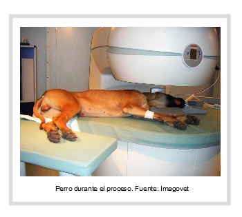
- The animal is placed under general anaesthetic and intubated, and the anaesthesia is maintained using gases.
The animal’s “constants” will be monitored continuously by a veterinary anaesthetist.
- The animal is placed on the stretcher and the study begins.
If the suspected diagnosis deems it necessary, a contrast agent will be injected intravenously to obtain more appropriate images.
- If necessary, when the procedure is over but the dog is still anaesthetised, it is possible to take a cerebrospinal fluid sample to study immediately, and to send a sample to the laboratory for subsequent examinations.
- The animal is taken to a suitable area to come round from the anaesthesia.
How much does an MRI scan cost?
When you ask for a quote for an MRI scan, you must be aware that you are not only paying to obtain some images, you are also going to a diagnostic services department where a team of human specialists will make sure that the procedure is carried out under general anaesthetic, under the safest conditions possible.
Only specialists in the area are capable of extracting the results from the images that need to be analysed. In some cases, the final interpretation of the results is provided by vets that have a qualification in telematics, so it may take a little longer to get the final results.
The price depends on the number of areas that need to be examined, the size of the animal and the additional tests that need to be carried out. In any case, the vet that makes the referral will send you to the diagnostic services that he/she considers most suitable.
For these reasons, the price can vary quite a lot, but as a guide, the price can oscillate between 350 and 800 euros.
Ortocanis Technical Team