A simple and brief explanation of the physical principles:
This imaging technique is the most widely used, it’s noninvasive and doesn’t require any preparation of the patient.
To get a radiograph of a patient, an X-ray machine is used. This device emits a beam of X-rays in the direction of the patient.
Part of the radiation is absorbed, another part is dispersed and the other manages to penetrate the body and is impressed onto radiographic film, resulting in a two-dimensional image. Lead protectors used by the staff taking the X-ray protect against the dispersed radiation.
Depending on the tissue’s density, thickness and Rx hardness, some bodies will absorb more radiation than others. The concepts of opacity and transparency stem from these characteristics. We classify tissues on a scale that goes from radiolucent (the radiation easily penetrates) to radiopaque (very little absorption occurs or no radiation at all ends up penetrating).
X-rays act upon a photographic emulsion so once revealed and fixed, the image plate has a blackening effect, which is the base of the radiologic image.
On this basis we classify tissues from more radiolucent (black) to more radiopaque (white):
AIR > FAT > WATER > CALCIUM (bone) > METAL > SOFT TISSUE > CONTRAST > ENAMEL
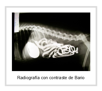
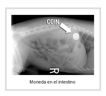
In the following images we can see materials that are completely radiopaque (metal and contrast): a coin in the stomach and a Barium contrast in a dog. The Barium is a contrast substance that the animal ingests in order to see its intestinal track marked clearly.
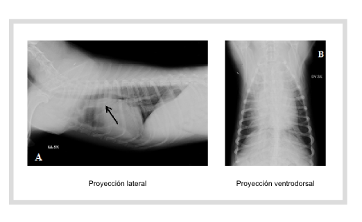
As the radiograph is a two-dimensional image, at least 2 perpendicular projections for each radiograph will ALWAYS be necessary in order to get a complete evaluation. This means that, for example, to do a radiographic study of the chest we will need to take a lateral and ventral-dorsal x-ray.
X-ray indications:
Your vet will know how a normal radiograph should look and will be able to detect pathological changes.
It is used to assess the:
-Cranium: jaw, sinuses, maxillary, tympanic bullae ...
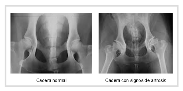
-Osteoarticular system: fractures, inflammatory and degenerative reactions of the bones (osteoarthritis), dysplasia, dislocations, neoplasms, reduced bone densities.
-Chest: ribs, pleuralspace, cardiac silhouette and the size of the large vessels (arteries and veins), changes in lung parenchyma, density changes due to pneumonia, accumulation of fluid (edema), tumors ...

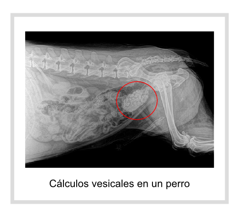
-Abdomen: we can evaluate the position and size of the abdominal organs, presence of intestinal obstructions, neoplasms, kidney or bladder stones...
In recent years, many veterinary clinics have left behind the conventional X-ray to use digital radiology, which speeds up the process, improves image quality and eliminates the generated wastes (revealing liquids and image plates).
While the X-ray is still a very valuable tool, sometimes it needs to be supplemented with other techniques in order to reach the final diagnosis.
For example, an X-ray may show an enlarged prostate, but an ultrasound will be needed to evaluate the characteristics of the tissue (cists, tumors.
An X-ray of the spine may show normal or a slight decrease in the intervertebral space, however an MRI will be needed to assess whether or not a disc is compressing the spinal cord.
In conclusion, each diagnostic imaging technique has its uses and limitations, some cover the limitations of others, and as such in many cases we need to use several of them in order to reach a definitive diagnosis.
Ortocanis Technical Team
![]()