Tag Archive for: orthopedics

Canine osteoarthritis is a very common disease in dogs. It appears as a result of the inevitable evolution of a joint that ages or becomes increasingly fragile due to trauma or malformation. This is a very painful condition that needs to be treated right away. It can affect all the joints of the body, both those found in the anterior and posterior extremities, as well as those that form the spine. In the case of senior dogs, the most common is that this disorder affects several joints at once.

protector rodilla para perroThe articular surface is covered by a tissue called cartilage, which plays a role very similar to the shock absorbers found in cars. In addition, it prevents the underlying bone from deteriorating due to the repeated rubbing to which it is subjected by continuous movement. Osteoarthritis is characterized by a progressive destruction of this cartilage and by an abnormal bone proliferation at the edge of the articular surfaces known as osteophyte, also called ‘parrot beaks’ when they are located in the spine. The affected joints lose elasticity, cause pain and prevent the animal from moving normally.

Evolution of deterioration
As a rule, this ailment affects, first of all, the high joints of the limbs: hips and knees, shoulders and elbows. Symptoms are more or less important depending on the number of joints affected. However, there is an unequivocal sign that makes us intuit that the animal is affected. Lameness usually manifests itself when the dog gets up and starts up after remaining immobile for a long time.

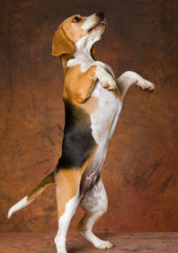
The pain prompts the animal to avoid support on the affected limb and, being impeded, stops running, and of course, jumping. As it evolves, the pain increases. When making certain movements, the dog emits small moans, it is even possible that the animal is irascible and tends to present aggressiveness when we try to manipulate the affected joint.

In more advanced cases, the joint may be partially blocked, making it impossible to perform certain movements. At this point, the animal hesitates to use the diseased limb. In addition, this lack of activity leads to a significant deterioration of the musculature surrounding the joint. As a result, the diseased area begins to atrophy, which increasingly complicates its use.

Two fundamental types
As a general rule we distinguish two types of osteoarthritis: primary and secondary. The first type usually affects elderly animals and appears due to the normal aging suffered by the joints due to the passage of time. In fact, it is a progressive and inevitable wear of the articular cartilages. Usually, this type of osteoarthritis affects different points at the same time.

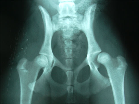
As far as secondary canine osteoarthritis is concerned, it appears as a consequence of a triggering factor, which causes the affected joint to stop functioning normally. For example, this type of osteoarthritis can appear due to trauma – a sprain, a fracture, etc. – or due to a birth malformation, such as hip dysplasia.

Another very common cause that causes the appearance of secondary osteoarthritis is obesity. If you do not control the diet your friend can have a weight well above the average that we find in the breed. The joints are not made to support such a significant overload of kilos, so they deteriorate easily. Unlike primary osteoarthritis, secondary osteoarthritis can affect animals of all ages and, as a general rule, usually affects only one joint.

Issue a diagnosis
The diagnosis of this disease can be based on three factors: the pathological history of the animal, gait examination and manipulation. When studying the history, the veterinarian must take into account old joint fractures, injuries that the animal has suffered a long time ago, as well as possible sprains. When observing the gait will look at whether the dog limps, even if only very slightly and incipiently, since lameness is an unequivocal symptom of the disease. Finally, when manipulating the affected area it is very possible for the animal to show signs of pain.

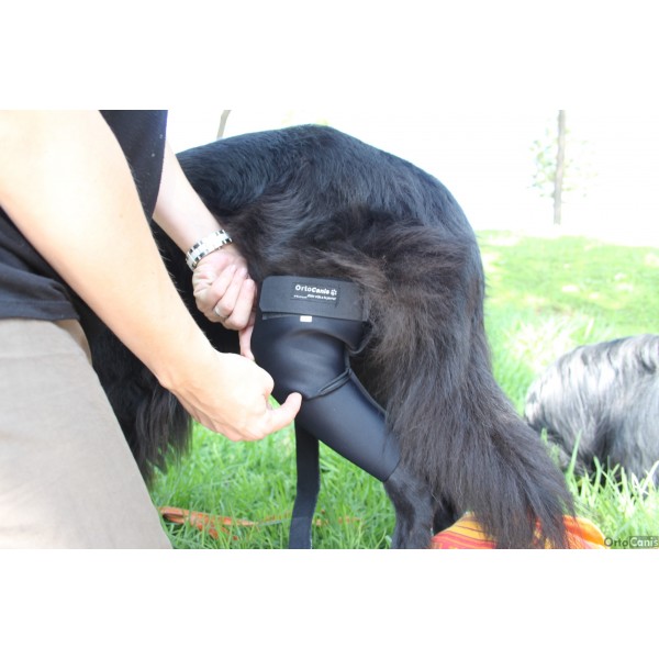
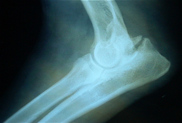
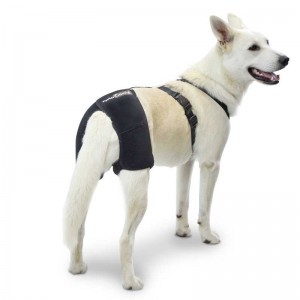
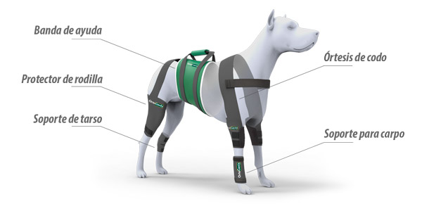
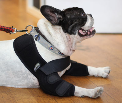
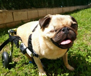
Often, the region in which the diseased joint is located is usually somewhat deformed due to osteophytes and muscle atrophy caused by the absence of physical activity. Many times a characteristic snap is detected when we move it. Through the radiological study, the animal health specialist will be able to determine the severity of osteoarthritis and establish the most appropriate treatment. Among the possible treatments, the appearance of specialized supports for some limbs that can effectively combat the deterioration of the animal’s quality of life stands out. The use of these supports is effective and has proven in countries such as the USA to be treatments as innovative as successful, however their use should be consulted with the veterinarian or animal physiotherapist.

Orthocanis Team

British scientists managed to reverse paralysis in dogs by injecting them with cells extracted from the lining of their own nose.

All of the animals in the study had suffered spinal cord injuries that prevented them from using their hind legs.

Researchers at the University of Cambridge, England, are cautiously optimistic that the technique may eventually play a role in treating human patients.

The study, funded by the Medical Research Council (MRC) and published in the neurology journal Brain, is the first to test transplantation in “real-life” wounds and not in laboratory animals.

Foreign scientists of the lining of the nose of animals are called olfactory enveloping glia (OEC) cells.

OLFACTORY ENVELOPING GLIA CELLS

The only part of the body where nerve fibers continue to grow in adults is in the olfactory system.

The enveloping cells of olfactory glia (OEC) are located at the back of the nasal cavity and surround the receptor neurons that allow us to smell and convert those signals in the brain.

Nerve cells need to be constantly replaced and this is promoted by the OCs.

For decades, scientists have weighed in that OECs could be useful in spinal cord repair. Initial trials with OEC in humans suggest that the procedure is safe.

These were cultured and reproduced for several weeks in the laboratory.

Of the 34 pet dogs that participated in the proof-of-concept trial, 23 received transplants of these cells at the site of injury and the rest received an injection with a neutral fluid.

Many of the dogs that had received the transplant showed considerable improvement and were able to walk on an exercise machine with the support of a harness.

None of the animals in the control group managed to reuse their hind legs.

Caution and optimism

The research was a collaboration between the MRC’s Centre for Regenerative Medicine and the University of Cambridge’s Veterinary School.

Professor Robin Franklin, a regenerative biologist at the STEM Cell Institute at the MRC and the Wellcome Trust and one of the report’s authors, said: “Our findings are extremely exciting because they show for the first time that transplanting these cell types into a severely damaged spinal cord can bring significant improvement.”

“We are hopeful that this technique can restore at least a small amount of movement in human patients with spinal cord injuries, but that is a far cry from the fact that they could recover all of their lost functions.”

Professor Franklin says the procedure could be used in conjunction with pharmacological treatments to promote nerve fibre regeneration and bioengineering to replace damaged neural networks.

Nariz
The animals received a transplant of olfactory cells.

The researchers claim that the transplanted cells regenerated nerve fibers throughout the damaged region of the spinal cord.

This allowed the dogs to regain use of their hind legs and coordinate movements on their front legs.

But no new long-distance nerve connections like those required to connect the brain to the spinal cord were developed.

According to MRC scientists in humans this would be vital for a spinal cord injury patient who has lost sexual and bowel function and bladder control.

Professor Goeffrey Raisman, Chair of Neural Regeneration at the University of London, who discovered olfactory enveloping glia cells in 1985, says: “This is not a cure for spinal cord injuries in humans, which may still be a long way off.”

“This is not a cure for spinal cord injuries in humans, which may still be a long way off. But this is the most encouraging development in several years and is a significant step on the road to achieving it.”

Prof. Goeffrey Raisman


“But this is the most encouraging development in several years and is a significant step on the road to achieving it.”

“This procedure has allowed an injured dog to walk with its hind legs, but the range of much higher functions that are lost with a spinal injury, such as those of the hand, bladder function, temperature regulation, for example, are more complicated and still very distant.”

Jasper, a 10-year-old dachshund, is one of the animals that took part in the trial.

Owner May Hay told me that “before the treatment we had to transport Jasper in a car because his hind legs were useless. Now he walks all over the house and garden and can keep up with other dogs. It’s wonderful.”

Source: BBC

Elbow dysplasia is a very common degenerative disease in young dogs. The elbow of dogs is one of the most congruent and stable joints of the body, allowing, due to its complexity, two axes or degrees of supination-pronation movement of the forearm and flexion-extension. Its complexity is given by its composition: humeroradial joint, humeroulnar and, proximal radioulnar.

Elbow dysplasia was initially used to describe the non-union of the anconeal process (AUP). Currently, osteochondritis dissecans (OCD) of the medial condyle of the humerus, the fragment of the coronoid process (FPC) and, the incongruence of the elbow (INC) are also included within this term. When one of these ossification defects occurs in an elbow, inflammation originates and over time an osteoarthritis is triggered in which cartilage degeneration occurs; for that reason, all these conditions are commonly associated with osteoathrosis of this joint and are an important cause of pain and claudication of the forelimbs in large and giant breed dogs such as the German Shepherd, Labrador, St. Bernard, Rottweiler, Neapolitan Mastiff, among others.

Of genetic origin multifactorial, especially in OCD and FPC. It affects males more than females and can occur uni- or bilaterally. The genetic component is the one that has the greatest influence although, the appearance of this pathology can also occur due to food, weight, environment, quality of ligaments, a lot of physical exercise or trauma.

The first symptoms may occur at 4-5 months when the dog shows exercise intolerance, lameness when starting a movement or after prolonged exercise. There are dogs that do not show signs of affection in the elbow until advanced ages where the process of osteoarthritis is very evolved. Others manage to maintain a normal degree of activity throughout their lives.

The fact of making a premature radiological diagnosis makes it possible to establish an adequate treatment and avoids the formation of osteoarthritis that produces pain and functional limitation of the elbow throughout the life of the animal. The diagnosis can be complemented with diagnostic tests such as CT or MRI

The evolution depends on the degree and type of injury, but it is usually unfavorable without surgery. Surgical treatment is good if degenerative changes in the joint have not yet occurred. In any case it is necessary to perform a good rehabilitation in order to:

  • Speed up the recovery process
  • Eliminate pain and inflammation
  • Decrease lameness
  • Maintain and/or improve range of motion
  • Maintain muscle tone, mass and strength
  • Minimize or slow down the effects of joint degeneration – osteoarthritis
  • Avoid compensation at the level of the neck, spine and extremities
  • Give the maximum capacities so that the animal is functional and that it, with a good quality of life

Physiotherapy treatment varies depending on the animal and the state of the lesion. It is important to start as soon as possible with the treatment so that it is effective and, to avoid drying them as reduced mobility and / or chronic pain.

The animal goes through different phases until its full recovery. It is essential to gradually achieve the objectives set. The recovery process is terminated when the animal is able to perform daily activities.

During the first three days after the intervention, it is important to act on inflammation and pain and prevent muscle atrophy and decrease in the joint arch from appearing. For this, passive techniques are used that reduce inflammation, produce analgesia and help maintain tone, mass and the arc of mobility. Among these techniques there are electrotherapy (segmental TENS and muscle electrostimulation), massage, passive mobilizations and cryotherapy (cold).

In older dogs or dogs that have not been intervened, the objectives will be the same as in animals that have gone through an intervention. It is important to eliminate pain because, with pain you can not work.

It is important from the beginning to massage and move the affected elbow as long as there is no veterinary contraindication and, respecting in the case of fixation, the period of healing and union of the fixed parts. Massaging and moving the affected area and limb helps maintain mobility, prevents loss of mass and tone and works the proprioceptors.


A gentle mobilization combined with different massage techniques help decrease inflammation and reduce pain.

With TENSat the segmental level we can produce analgesia and decrease the amount of drugs administered. There are animals that have intolerance to certain drugs that produce analgesia and with TENS the pain can be reduced. TenS can also be used directly on the injured or operated area, as long as there is no osteosynthesis material underneath, since an internal burn could occur.

Muscle electrostimulationhelps prevent the onset of atrophy and maintain muscle mass and tone. With electrical stimuli we can stimulate nerve conduction.

At the beginning and end of the session the coldis used since it has properties that act on the decrease of the inflammatory response, edema and pain.

From the fourth day and during the next two weeks when the inflammation and pain have disappeared it is time to introduce simple active exercises such as shaking hands or small walks on a leash to force the animal to make an equal support with the four limbs and thus, prevent a decompensation between limbs from appearing due to not having a correct support on the ground. The walks is an exercise that increases the duration until full recovery.

Once the stitches have been removed, the animal can be introduced into the water. The advantages of water are used to improve recovery. Hydrotherapy (underwater treadmill) facilitates the station of the animal without loss of balance and, thanks to flotation, without having to support all its weight. In addition, flotation allows animals with bone pain and low muscle mass to work. The pressure of the water exerted on the body of the animal increases the sensitivity and decreases inflammations and edemas. The work in the water, underwater tapes or swimming increases as the animal recovers. In addition, with water, we can recover the motor pattern, increase mass, tone and strength, work on respiratory capacity and maintain and / or improve mobility.

Once the acute phase has passed 48-72 hours and without risk of infection or inflammation, heat can be introduced that helps to elastify the tissues, decreases pain and increases vascularization among others.

The use of boards, plates, balls and trampolines are important to work on balance, proprioception and above all the integration of the affected limb.

It is already in the last phase, from two weeks, when the dog has integrated the gait pattern, exercises are performed to improve the quality of movement. They are more complex active exercises to integrate the affected limb or limbs. With active and proprioception exercises it is possible to increase muscle tone, mass and strength; coordination and balance and range of motion are worked on. Rails with different surfaces, cones, bars, circuits, up and down stairs and ramps (staircase with inclined plane) are used.

Throughout the recovery treatment and in animals with developed osteoarthritis it is essential to reduce the weight on the joints of the elbows. For this purpose , special support harnesses for elbows are used. In addition to decreasing weight, pain is reduced and does not hinder movement, the animal feels more comfortable; the joint is protected at all times from chafing and blows and helps maintain the heat that the animal gives off, which leads to a relief of the affected area.

At home, special care should be taken for animals suffering from elbow conditions. This care is necessary during and after treatment:

  • Avoid slippery floors
  • Avoid ramps and stairs at the beginning of treatment in operated animals and in animals that do conservative treatment. Once rehabilitated, ramps can be used to help get on the sofa and the car, since it is recommended that they do not do it alone, there could be a recidivism.
  • It is recommended that they rest on soft and clean surfaces, but that they are firm enough to help the incorporation of the animal thermal mattress for dogs
  • Keep skin clean and dry skin
  • Use special plates at your height so as not to strain the elbow joints
  • Correct diet and weight control. Being overweight harms the joints and generates more pain for the animal

It is very important to create an exercise routine and environment to help keep the animal comfortable and with quality of life.

Orthocanis Team

1. What is hip dysplasia?

It is one of the most frequent diseases in dogs, especially large breeds.

2. What is the disease?

In a mismatch between the head of the femur and the acetabulum, that is, a bad gear of the hip joint. The femoral head is partially outside the acetabulum and the hip cannot work properly.

3. Does it affect some races more than others?

Yes, hip dysplasia practically does not exist in Greyhounds and yet it is very common in San Bernardo and Mastines. It is also quite common in the German Shepherd, the Golden Retriever and the Rottweiler among other breeds.

4. How to know the severity of dysplasia?

There are several degrees of dysplasia, but there are also different ways to measure it, the most common being an X-ray that should be done on dogs near the year of life. This x-ray must be performed in a very specific position and with the muscles relaxed, so it is usually necessary to anesthetize the dog.

5. What are the symptoms?

Normally lameness in dogs from 5 to 10 months but can remain without problems during youth and adulthood and debut with major problems in the geriatric period. Movement to the sides of the hips may also be indicative of hip dysplasia.

6. Does it have anything to do with osteoarthritis?

They are two different problems, but hip dysplasia, the mismatch of the joint, can cause over time that this joint degenerates and ends up suffering from osteoarthritis in the hip. Osteoarthritis is what hurts, especially in old age. Dysplasia may be more responsible for the dog’s development of osteoarthritis.

7. Is there treatment?

There are several treatments, various surgeries before the year of life and other methods such as hip prosthesis when the dog is older and has finished growing.

We also have palliative treatments such as anti-inflammatories or cartilage protectors (chondroprotectors). In very serious cases you can put a canine wheelchair and the dog continues to walk but without bearing the burden of its weight.

Ortocanis.com Technical Team

Electrostimulation for muscle potentiation

Electrostimulation is a very useful tool to enhance the muscles of a dog that has suffered an injury or surgical intervention and as a result has muscle atrophy.

We can use electric current to stimulate the muscles, enhance it and make it work, it is specifically indicated in cases of muscle atrophy, and especially in cases where the dog can not do active work. If the dog can do active work, electrotherapy will be a support, never the main source of work.

Form of the impulse that we will use will be the Rectangular, symmetrical biphasic the main authors indicate it: Thepaut Mathieu 1992, Kramer 1984, Bircan 2002

pack-tens-para-perros

Human electrostimulation apartments can be adapted to electrostimulate dogs without too many problems, but we must ensure that we can vary the frequencies, and especially the amplitudes of the electrical impulse to adapt well to the characteristics of the dog.
There are no good studies of Cronaxia in dogs, the approximate values are 0.3 human, 0.2 Horses and in dogs by studies of less depth intuit similar values;
Sawaya – Meallier 2006, Brodart 1998, Coarasa 1999, Ramon 2007
The Intensity according to Hultaman 1983, and Ogino 2002 should be elevated with visible contraction, without reaching the limit of pain but discomfort. The dog must comfortably withstand the session but the muscles must work intensely.

The frequency will be marked according to the objective, various authors validate this option: Pougheon 1992, Busko 1989, Vanderthommen 2002.

Objective Frequency Treatment time Standby time
Relaxation: 5 Hz Continuous 0
Heating: 5 Hz Continuous 0
“Endurance”: 10-20 Hz 9 2
Atrophy: 33 Hz 6 6
Strength: 50- 100 Hz 5 25
Explosive force: 100-200 Hz 3 30

The parameters may vary slightly according to individual cases.
Rest time can be adjusted if it is active, it is advisable to use ramps before strong muscle contraction.
The intensity should always be the maximum with relative comfort.
Total time between 10 and 30 minutes depending on the phase of the injury, fatigue the muscles excessively does not help to enhance the muscles correctly.

It is important to have a device that meets all the requirements, if it is going to be used by a canine rehabilitation center or veterinary hospital, a more professional desktop apartment is recommended and with it we can use specific programs for each of our patients.

The use of rubber electrodes and contact gel is equally recommended in animals with hair.


Orthocanis Veterinary Team

Baner silla 728x90

Cold is one of the most used elements for the treatment and prevention of muscle pain and joint or soft tissue inflammations.

After surgery or in cases of soft tissue injuries such as tendonitis or muscle injuries, the application of cold reduces pain, prevents the formation of excess inflammation and helps resolve it if it has appeared.

bolsas-de-agua-caliente-o-hieloThere are several studies that talk about the effectiveness of the use of cryotherapy in its different aspects, application of ice, single-use bags of instant cold , cold pack, neoprene devices for use with cold packs or crushed ice, immersion in cold water with ice cubes …

All of them have their advantages and disadvantages.

Techniques that use ice directly, or immersion in cold water have the problem of wetting the skin of the animal and transmitting too much moisture to the body of the animal, in addition the ice water that is just touching the skin of the animal can produce burns and irritations, in human it is always recommended to put a cloth between the skin of the patient and the ice.

Sometimes, in dogs we think that their own coat is enough, but we can have problems with the water, very cold, which is just touching the dog’s skin. Therefore it is better to avoid this direct contact.

There are very expensive “dry ice” equipment that are machines that emit very cold air and are used in human rehabilitation centers, especially in sports centers, they clearly lower the temperature of the tissue if they wet the skin and without creating moisture or irritation. The dog’s hair can be an impediment to the penetration of the cold of this type of devices, this factor together with the high price make this technique practically not used in dogs.

The fastening system is another important factor; exist in the marketbolsa-de-frio-instantaneovery well designed restraint systems that allow the dog up to a certain mobility without the ice falling, it is extremely advisable to use straps or other more evolved fastening systems since this way we ensure the intimate contact of the ice with the dog’s skin and that this contact will last as long as the treatment lasts.

The estimated treatment time should be between 15 and 20 minutes, the most recent studies recommend these times since they are the ones that allow to maintain for a certain time a more or less deep tissue, such as a joint capsule of the knee at a temperature considered therapeutic of 15ºC. Precisely the knee is one of the joints where more ice can be used as a treatment system and a Device that fixes the ice well to the knee can help us a lot to be more effective. If the internal tissue does not fall to 15ºC we are not taking advantage of the possibilities of cryotherapy.

The latest novelty in cryotherapy treatment is to agree with compression and the latest evolution of compression is that it is alternating. There is only one machine on the market that provides these two components, cryotherapy and intermittent compression in a single device, the cold and compression system.

Ortocanis technical team

Baner silla 728x90

It is one of the most used instruments in physiotherapy. It is a device that emits acoustic waves of much higher frequencies than those audible by humans. The frequency used as a therapeutic tool is 1×10⁶ Hertz, i.e. 1 Mega-Hertz (MHz), so they are not audible by any mammal.
Usually in veterinary clinics and hospitals, ultrasound is used for ultrasound scans that use the same type of wave. The difference is power, frequency and application time.
In therapeutics we use frequencies of 1MHz for deep treatments, up to 8 cm and frequencies of 3MHz for more superficial problems. The power ranges between 0.2 and 3 Watts / square centimeter.

Effects on tissues:

The main effect of ultrasound on tissues is anti-inflammatory. We usually use it in tendons, joints or inflamed muscles; it has excellent results in both acute injuries and chronic injuries, although we must adjust the powers.

The analgesic effect is another of the most sought after in rehabilitation, normally when we deflate a structure we manage to reduce the pressure in the nociceptors that are the receptors in the body that send the painful signals, by reducing the pressure in these receptors, we reduce their stimulation and therefore decrease the intensity of the signals they send until they disappear. If there is no sign of pain, there is no perception of it.

soporte para perro con displasia de caderaWhen we have a fibrosis in the different soft tissues: muscles, tendons or ligaments, we can apply continuous and then pulsating ultrasound at maximum power. This way we will find a good defibrosing effect.

Another of the classic applications of ultrasound is the application in muscle contractures, with ultrasound we can reduce and even eliminate them.

Continuous ultrasound generates heat by the vibration of the molecules and both the pulsating and the continuous increase the permeability of the membrane, which is what favors together with the mobilization of the molecules the anti-inflammatory effect.

Application:

Ultrasound must be applied by moving the head all the time that the treatment lasts, either making small circles or following the direction of the treated tissues rectilinearly. If we do not do so, especially in continuous mode, we can damage tissues and produce significant burns.
It is necessary to use a means of contact, either contact gel or directly in direct immersion (sub-aquatic ultrasound).
It is also important to take into account the hair of the animal, this hinders the transmission of ultrasound and therefore it is very interesting to be able to shave the dog before applying the ultrasound sessionperro-multimedia-600x300_6

Approximate time between 5 and 15 minutes
Power between 0.2 and 3 Wats/cm2
There are different head measurements depending on the needs.

Directions:

Ultrasounds can be used in any pathology of the dog that occurs with joint or soft tissue pain, such as tendonitis, bursitis, arthritis, bruises or major bruises.

We can also use ultrasound in chronic problems such as hip dysplasia, elbow dysplasia, knee osteoarthritis, or hip osteoarthritis.

All surgical interventions produce an inflammation of the tissues that have been operated, ultrasound is a very good tool to control inflammation and post-surgical problems such as rupture of the cranial cruciate ligament, patella dislocation or others.

Ortocanis technical team

Baner silla 728x90


Owners often ask themselves questions about whether to shelter their pets in winter, this question at the same time comes with many others of this type:

Don’t dogs already have enough hair?
Is it a bit ridiculous?
Does it depend on the breed?
Does it depend on the area?

Is it normal in small dogs that are colder and silly in large breed dogs and especially with long hair?

All these questions may have more than one answer, but in general we SHOULD shelter dogs in winter, especially if we are in cold areas.

Older dogs are more likely to suffer from cold than younger dogs, who have more vitality. Older dogs especially with problems such as osteoarthritis suffer a lot with the drop in temperatures, we must do something to protect dogs with osteoarthritis from cold weather.
Dogs small breeds are more prone to cold than large ones, so they should be warmer.
There are some large breeds that are very prone to suffer cold and have a very bad time especially very short-haired dogs such as greyhounds.
The amount of hair influences, we will not shelter a Nordic dog in a Mediterranean city, but perhaps yes, if we go to the snow at sub-zero temperatures and do not do physical activity.

Regarding the ridicule is very opinionable, I could be more embarrassed to see the dog shiver and do nothing to help him.abrigo-para-perro
Elderly dogs are very grateful for a blanket in winter, and even in warmer times if they suffer from osteoarthritis in the spine or hips, the typical hip dysplasias of Golden, German Shepherd … they improve a lot with special thermal blankets. In winter especially if the dogs live in cold areas, or very humid or mountain, when we do not want the dogs to get wet on their outings, we can use waterproof blankets that also protect the muscles and joints from the inclemencies of the weather.

In summary, if we have a small breed dog or a large breed with short hair, or an elderly animal in any of its breeds and sizes , we must shelter it in winter.


Ortocanis technical team

Baner silla 728x90

Hip dysplasia Irresponsible breeders, feeding, environment?

Currently, with only three months of age it is possible to know the existence of small anomalies in the conformation of the hip/femur joint, which will lead to dysplasia.

Origin of hip dysplasia in dogs:

In colloquial language (today we avoid veterinary terms) hip dysplasia is a “failure” in the head joint of the femur-hip. If the head of the femur is not lodged in the hip perfectly, there is a deterioration of the cartilage that protects the joint, and that deterioration is degenerative and irreversible. But why does hip dysplasia occur?
Genetic inheritance. Hip dysplasia is inherited, and if breeders do not perform the necessary tests to know that their dogs are free (certified X-ray) and that previous generations too, puppies can suffer from it. Many breeders (and more particular) ignore these x-rays (eye, there is no breed safe from the disease). Hopefully they include a clause in the sales contract in which they will give you a puppy if you prove that the one you have bought has dysplasia (as if they were appliances).
Environmental factors. In the period of growth (until the year, but especially critical the first six months of life), slippery floors, sudden exercises, jumps … Puppies with a limit hip can aggravate their situation if care is not taken in these critical months, and vice versa, they will be able to lead a perfectly normal life if they develop correctly in these months (even if their hips are not perfect).
Feeding. The months in which dysplasia develops are those of growth, and the slower the puppy grows the better. Foods very high in protein have been linked to the onset of dysplasia. Chondroprodectors help during growth (in predisposed individuals or breeds, always under veterinary supervision).
About prevention in line with the above, if the breeder is responsible and has all the controls done we still can not sing victory. It is very important that the puppy has a good diet according to his growth needs, which does not get fat (the image we all have of a rolly puppy is typical, but not healthy), supported by chondroprotectors if necessary, that the exercise is restrained (avoiding strange movements, and especially jumps and forced postures of the back three), be careful with the floors of the house (if they are slippery it is not a bad idea to get some old carpets that last us a few months).

Some exercises and “tricks” are very demanding with the hip, and therefore dangerous in puppies and young dogs.

And the greatest prevention: radiography There are many puppies that can limp for causes that have nothing to do with dysplasia, and in the same way, there are asymptomatic with serious femur and hip problems. The plate is painless, economical, and the only truly reliable method. At present we can know the state of the hips of our puppy from as early as three months (PennHip method), so that conservative treatments can be established, or in the case of an intervention being necessary, which is not drastic but reconstructive, preserving the joint. Until the year of age it is not possible to ensure that the hip has had a perfect development and, therefore, it will not be until then when the dog can start in canine sports (“start” is to go little by little) performing more demanding exercises with his body.

Source: www.doogweb.es

www.ortocanis.com


Ortesis de codo para perros con higromas o callosThe five keys to having a healthy and happy dog at home and enjoy it

There are different aspects that are important when we have a dog as a pet at home. Having a healthy pet means not only having it in good health but also other aspects.

The care of the health of the pet, hygiene and a correct education of the dog are the three pillars of the good coexistence of dogs and people.

The health of the pet is an aspect of vital importance, a healthy dog can bring us problems but a sick dog or with a weak state of health will surely bring them to us.

Healthy Habits are essential to maintain the good health of our pets:

  • Routine veterinary visits and vaccinations
  • Regular physical activity

duc-i-ana-2If we complement it with a correct hygiene (also important for health) and a careful education of the dog important for the family-dog relationship, we will have a set of aspects that will greatly improve the relationship with our dog, its quality of life and that of the whole family.

The correct feeding:

Dogs must eat feed, many times we wonder if it would not be better for dogs to eat homemade food, or if they can live well with what is left over from a house, as it was done before. The answer is always the same, canine feed is a complete, balanced food, formulated by veterinary nutritionists who determine the needs of each dog according to their breed, age and physical activity and develop the most balanced and complete diet.

Canine feed, as long as we buy the type that best suits our animal, will be the best food we can give our pet. We must respect the measures recommended by the same brand or ask for help from our veterinarian, he will be the most suitable to explain the amount of feed you should eat and in cases where necessary if we can complement it with a special dietary supplement.

Routine visits and vaccinations:

Not only for our pet but also for everyone else in the neighborhood, even for our own safety and those of the people who live with us or our neighbors it is extremely important that we keep the dog’s vaccination card up to date.

Proper vaccination prevents diseases that can become very serious for our animals.

Hygiene:

It is another important aspect, although sometimes forgotten about pets, hygiene not only means taking the dog clean, but taking care of its skin, its hooves, its mouth and therefore its breath, and that it has the appropriate place to relieve itself without staining and where we can pick them up and deposit them in the appropriate place.

Depending on the type of dog, the breed, its physical activity we must wash it more or less, and in certain breeds we must have special attention to specific points. For example, Cockers have a special tendency to make recurrent otitis, even chronic, this is because due to the position of their ears and their size, the ears breathe very little and it is an excellent breeding ground for bacteria and fungi; it is avoided with a simple spray every so often and a correct washing of the dog’s ears much more commonly than is necessary in other breeds.PastedGraphic-1

It is good to be informed of these aspects when we adopt or incorporate a new member into the family. You can consult in many forums specific to dog breeds, kennels, dog clubs and of course whenever you have doubts to the Veterinarian.

Physical activity:

It is the great forgotten in Spanish dogs, I feel envious when I see in American movies the dogs doing physical activity with their owners in Central Park …

Some privileged Spanish dogs go for a walk three or four times a day, but they go for a walk to relieve themselves and especially in winter these walks are limited almost exclusively to this activity. It’s not that it’s wrong to take the dog out to urinate or defecate, the problem is that too many dogs ONLY come out to urinate and defecate.

The walk should be an important part of the owner’s relationship with his animal, the dog should feel free, satisfied, happy with the walk and should serve for him to do physical activity. Therefore it is important that one or two of the walks that the dog takes throughout the day is much longer and the dog, especially if it is young, can run freely, jump, play with other dogs …

Education:

Another aspect in which, as a whole, although it hurts to recognize it, we have been a few years behind Western Europe and the United States in Spain, even with certain Latin American countries, is the education of our dogs.

Many times we complain that they do not let us enter hotels, restaurants, shops or cafes with our dog. It is true that we can oppose many problems if we look for a hotel where pets are accepted in certain Spanish cities. On the other hand, many of us have traveled to Holland, Belgium or of course the United Kingdom and have seen small dogs on planes, and larger dogs in Perro en silla de ruedas
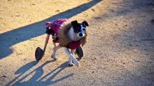
cafes and restaurants. We have seen them but we have not heard them, nor have we smelled them nor have we noticed their presence until the owners of the animal have left the premises. The dog was missing the entire time he was in the restaurant. Nothing like the dogs that you can find in beach bars, which is one of the few places where they accept them, and not always, competing for the neighbor’s croquette or for the piece of ham that has fallen to the lady or for directly the grilled cuttlefish that have brought you and smell wonderful.

We must sing a collective mea culpa about the education of the Spanish canine cabin, to more and better education more and better respect and understanding on the part of people who do not have pets. A dog that does not bother anyone would not count anyone and few people oppose its presence. A dirty dog, with a foul breath when he throws it directly to the face of the neighbor’s baby who in his stroller filling it with drool and scaring the child is normal to provoke the upset reaction of the child’s parents.

Orthocanis Veterinary Team

www.ortocanis.com

Ayudas para perros con artrosis