Tag Archive for: orthopedics

Improving the muscles of the hind limbs in dogs is an interesting option both in dogs that are being rehabilitated from problems in the hind limbs and in dogs that are initially healthy, but that have weakness in the hind limbs.

The first thing we have to do is evaluate the patient. Observe the muscle mass of the posterior ones, palpate this musculature, look if apart from muscle atrophy there is tension or flaccidity.

If we observe muscle tension it will be convenient to start the rehabilitation by unloading the muscles, with massage, with TENS, with heat … on the other hand, if we observe weakness and flaccidity, we must begin rehabilitation with Electrostimulation or light muscle toning exercises.

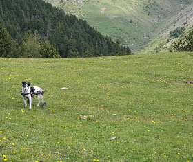
Walks is the easiest way to start a rehabilitation, it should always be pain-free, if the dog has pain you should go to the veterinarian and take the necessary pharmacological measures to resolve the situation, a TENS can also be very useful to eliminate pain during walks.

Another important point is the way the dog walks, if it mainly uses the forelimbs to perform its gait this exercise will not bring us practically any improvement in the strength of the posterior ones.

arnes-de-soporte-para-perro
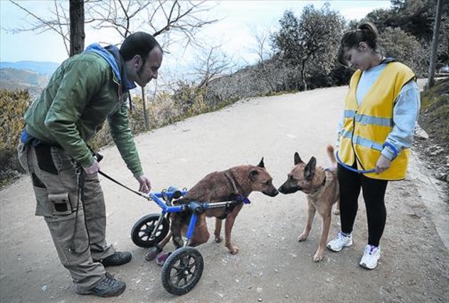
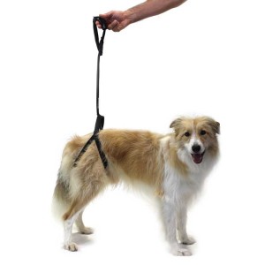
It is important to get the dog to work with the hind legs, this can be achieved by walking on the short leash and indicating to the dog that he must carry weight in the posterior ones, it will be practically impossible if the dog has pain in the hips, knees or some other structure of the posterior third.

Squats (making the dog sit up and get up a few repetitions in a row) is going to be another of the exercises we will use; we must reserve this exercise for when the dog does not have pain in the subsequent ones and his muscle mass is enough to withstand this exercise or failing that we can help ourselves with a harness for later to help lift the dog.

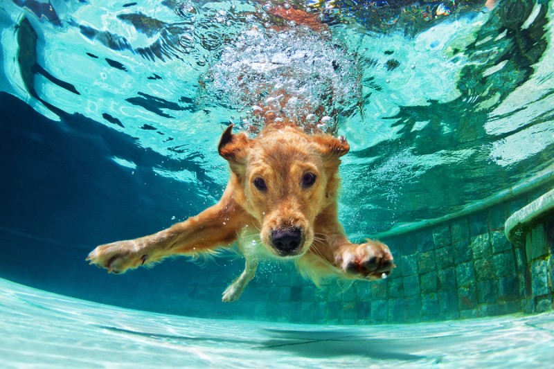
Working in the water with the underwater treadmill is one of the best exercises since they strengthen the muscles protecting the joints. The sloping walks is another of the exercises that we must practice, the inclination of the slope will force more the posterior third and the dog will work more and increase the muscular power of the posterior third. This can be done both on a climb and on a rolling tapestry with inclination.

soporte para perro con displasia de cadera

In the later phases of treatment weights can be used for muscle strengthening in dogs.

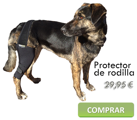
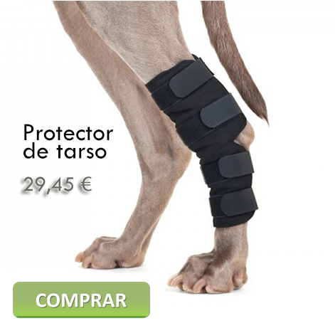
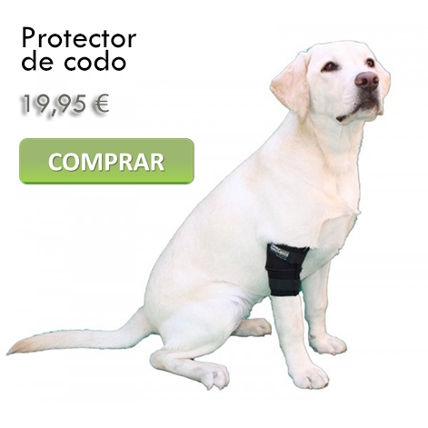
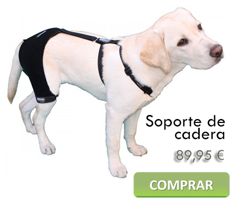
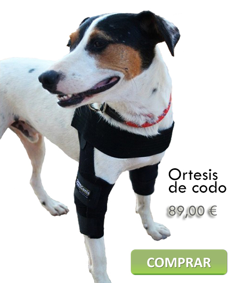
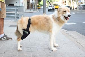
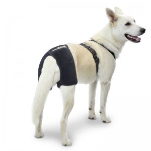
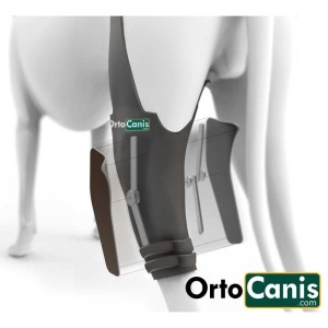
There are several solutions that can help us to be able to perform these exercises better, that the dog feels better and can correctly perform these exercises thus increasing the strength in the posterior third such as the knee pads, tarsus supports, hip support, or boots or foot protectors in case of having problems in the plantar area.

Orthocanis technical team.

www.ortocanis.com

Baner silla 728x90

There is talk of patellar dislocation in dogs, when the patella, a small bone located in front of the knee joint and whose correct position is necessary for a good functioning of the animal’s limb, gets out of its place, trochlea, causing functional pain and weakness in the dog.

Within the dislocations, the medial dislocation is the most frequent. It arises in 80% of cases while the lateral only occurs in 20%. Between 30% and 50% of cases are bilateral and more frequent in females than in males, especially small breeds and toys.protector-rodilla-canina-perro articulada

Lateral dislocations can occur in adult small breeds and in large and giant breed puppies.

It is a pathology that is characterized by a poor alignment of the limb, deformations occur during the development of the animal, which cause the patella to come out of its place. It may be due to a congenital disease or, in some cases, caused by trauma.

It is convenient that dogs suffering from this congenital pathology are not used in reproduction since it is transmitted through generations.

There are dogs that have patella luxation due to trauma. In these cases, the dislocation is generally associated with a rupture of the anterior cruciate ligament of the knee.

Depending on the clinical signs and later on the radiological results, dislocations can be classified into 4 degrees:

patellar lux_f
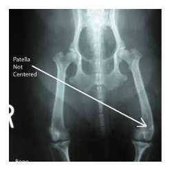
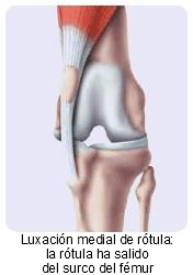
Grade I – Intermittent patellar dislocation causing limb lameness when out of place. In the dynamic test, every three or four steps they raise their leg by flexing the knee or they take a little jump.

Grade II – Dislocation that occurs more frequently than Grade I. The patella dislocates easily. There is a slight external rotation of the leg. Many dogs live with this grade for years before progressive arthritis manifests as lameness or more serious causes.

Grade III and IV – The patella is permanently dislocated, with very noticeable external rotation of the leg. There is moderate lameness. If it is bilateral, dogs walk with bowed legs, turning the feet inward and bearing weight on the forelimbs. In the most severe cases, it can be confused with hip problems.

In addition, the animal presents pain, crepitus and increased sensitivity in the knee, which leads to it reducing its activity, even refusing to go up and down stairs, the car or the sofa.

Treatment depends on the degree of dislocation and lameness, but in most cases surgical treatment consisting of soft tissue repair, bone reconstruction, or a combination of the two is required. There are infinite techniques and the orthopedic veterinarian chooses the most suitable one(s) in each case.

luxación de rótula

Among the most used techniques we find: Overlay of the medial or lateral retinaculum, overlay of the fascia lata, anti-rotational suture of the patellar and tibial ligaments, desmotomy/capsulectomy, quadriceps release, trochleoplasty (trochlear chondroplasty, sulcoplasty resection, trochlear sulcoplasty), transposition of the tibial tuberosity, patellectomy, osteotomy…

As a novelty, orthoses are being applied for knee dislocation as a means of conservative orthopedic treatment that allows the patella to be kept within the femoral condyles and avoid pain and instability. These orthoses are made to measure and are very useful in cases where surgery has failed, you cannot or do not want to operate for different reasons.

Marta Subirats & Toni Ramon

animal physiotherapists

Ortocanis technical team

Baner silla 728x90

Do you remember Glory, a dog with only one leg? We already published a post about Glory the dog with just one paw in case you want to know the beginning of the story.

Glory got her three prostheses thanks to anonymous donations. But its use always requires a period of adaptation and for Glory it was the first contact with an orthopedic device, imagine wearing no less than three of them … Here is the video of his first steps with prostheses:

As you can see, at first it looks weird and it is difficult to control the steps. But with a good motivation like food he manages to forget those sensations and starts walking

Glory has been impressing owners and veterinarians with her ability to adapt to something so difficult. The rear prosthesis resisted a little more, but nothing is insurmountable. This is Glory 16 days after his first contact with the prostheses:

Adapting dogs to prostheses is not done overnight, but Glory has shown that dogs are capable of this and much more. She has been the first to demonstrate it and has opened the door to this type of procedure, which allows to give a more than dignified life to dogs that until now were destined for immobility or euthanasia.

Bravo for Glory and her caregivers!

Thank you to everyone who made it possible.


Férulas caninas


Ayudas ortopedicas para perros

New technical and orthopedic aids for dogs are used in a wide variety of cases. These increase the mobility of dogs by providing the necessary support to maintain daily activity. Dogs need to exercise to maintain their physical and emotional well-being. If a dog is unable to exercise, it can develop problems such as heart disease, obesity, bone alterations, muscle atrophy, and emotional problems such as aggression or anxiety.

Applicable cases

Technical aids can be used for sprains, muscle weakness, arthritis, hip dysplasia, post-operative rehabilitation, lameness, weakness or pain in the joints that end up generating a mobility problem. The technical aids should allow the dog to perform its daily activities, but they should also be comfortable and offer protection against irritations and ulcers on the dog’s skin so it is important that they are performed by specialists. We list the most frequent:

Knee

Protector de rodilla canina

Knee protectors are typically used after surgery for rehabilitation of the anterior cruciate ligament (ACL), patella dislocation, medial collateral ligament (MCL), lateral collateral ligament (LCL), or posterior cruciate ligaments (PCL). Knee pads provide support and stability, prevent muscle loss during the recovery period, and reduce the risk of further injury. Knee protectors are also often used in cases where surgery is not performed.

Tarsus and Carpus

Protector de tarsoSoporte para carpo


Tarsal protectors are indicated in cases of osteoarthritis but also for post-surgical rehabilitation or injury to the Achilles tendon, nerve damage to the hind limb or foot and rehabilitation of the hock joint. They can also be used as long-term support in non-surgical conditions. A tarsus or hock protector stabilizes the bone of the back leg, as well as the muscles and tendons of the limb and knee. It works similarly to a knee brace in humans, allowing the dog to move without straining the joint. It also prevents the dog from re-injuring the affected joint or ligaments.

Elbows

Protector de codo canino

An elbow protector is used to support the elbow joint after surgery or to reduce pain from arthritis, degenerative joint diseases, or other similar conditions. It provides control in lateral movement while allowing for normal extension and flexion while treating and protecting hygromas and calluses.


Soporte de cadera caninoHip

A hip support holds and increases the temperature of the dog’s lower back and hip. Made of special neoprene, it surrounds the lower back, hip and upper leg and is fastened thanks to a Roman-type harness. Hip supports are commonly used for dogs with mild to moderate hip dysplasia and localized osteoarthritis.

Shoulders

Ortesis de codo canina

Medial shoulder instability (MSI) is one of the causes of lameness in the front legs. A special support would allow the shoulder to be supported, which would limit the extension, flexion and abduction, allowing the dog to support its weight. These aids can be used after surgery or as a non-surgical treatment but also preventively.

Physiotherapy as an alternative treatment in compression of Cauda Equina


SUMMARY

The cauda equina, is the set of nerves and nerve roots that originate from L6 – L7 innervating the posterior train by means of the nerves: Sciatic, Obturator, Pudendo, Femoral and Pelvic.

When generating a trauma at this level, mild to severe compressions can occur generating in the patient pain, claudication of hind limbs, contracture of all muscle mass, proprioceptive deficit, ataxic gait, incontinence, among other clinical signs that will guide the Veterinarian to establish an adequate medical treatment and in the case of the physiotherapist, locate the areas of pain and improve weight support, resistance, bodybuilding (avoiding muscle atrophy as much as possible), joint mobility and proprioception.

The diagnosis is made by radiological study and magnetic resonance imaging of the spine at the level of the lumbo-sacral joint, treatment options

consist of: cage rest, allopathic (NSAIDs, analgesics, corticosteroids), homotoxicological, physiotherapy and rehabilitation, being an excellent option the synergy between homotoxicological treatment and physiotherapy (integral analgesia).

This paper presents the case of a patient of a Pinscher, female, 10 months, which is taken to consultation after having suffered a trauma by fall presenting as main signs paraparesis, contracture of all muscle mass in hind limbs, proprioceptive deficit and ataxic gait. It was decided to perform physiotherapy and rehabilitation without suspending the medical treatment established from the beginning, managing to reduce the recovery time, improvement in the patient’s condition, elimination of the cause of the physical alteration, pain relief, reduce inflammation and resumption to daily life.

Keywords: cauda equine compression, physiotherapy and rehabilitation.

Interested? Here’s the full article


Angelica B. Ortega Vasquez

Ortocanis.com Collaborator


Hip dysplasia is a very common problem in certain breeds: BullDog, Bordeaux Doge, St. Bernard, Neapolitan Mastiff, German Shepherd, Rottweiler, Golden… all of them have an incidence above 20%.

Dysplasia is a multifactorial, multigenic and hereditary disease, that is, there are several factors that predispose and cause hip dysplasia, there are several genes involved in its appearance and it has a hereditary character.

Environmental factors are becoming more and more important in the development of hip dysplasia, the genetic factor is necessary to develop the disease but this is not the only factor. Genetics is a necessary but not exclusive factor, that is, you can have the genetic predisposition and not develop the disease but if you do not have a predisposition it is sure that it does not develop.

There are several degrees of dysplasia, and also those that appear when the dog is a puppy or those that give problem already in adulthood; but in this article we will focus on the treatment and specifically on the treatment of dysplasia to young dogs.

Classification of the degrees of dysplasia according to the OFA :

Grade I: minimal alteration with small subluxation and few degenerative changes.

Grade II: marked lateral subluxation of the femoral head, 25-50% of which is outside the acetabulum.

Grade III: 50-75% of the femoral head is outside the acetabulum; there are important degenerative changes.

Grade IV: dislocation of the femoral head with flattening of the acetabular border and femoral head; there are major degenerative changes.

The presentation in the young dog is puppy hip x-ray normally between 5 and 6 months and is marked by a significant limp.

A dog is not considered to be free of dysplasia until at two years of age already completed no problems or inconsistencies are observed in the control x-rays.

Food is one of the factors that predisposes to the appearance of hip dysplasia, Calcium-Phosphorus imbalances that must keep a correlation Ca1.6% – P1.1% and above all not overfeeding or providing excess proteins allows us to minimize the incidence of hip dysplasia. A hypocaloric diet from 3 months to 8 months protects dogs with rapid growth from dysplasia. Excess weight at 60 days is another factor that predisposes to the disease.

Hip dysplasia in the puppy usually debuts from 5 or 6 months, before it is not possible to observe any problem and the dog has been completely normal and has developed normally. The debut is usually presented as a sharp limp that prevents the dog from playing as it had done to date. We can observe changes in the desire to play, negative when going for a walk, to relate to other dogs or owners. Mood swings, frequent slips of the hind legs, discomfort and even refusal to be touched and the fact of “fleeing” from children in dogs that until a few days ago were playful and affectionate are frequent.

Sometimes when you reach 90% of the growth between 8 and 11 months the signs can be reduced and even disappear. Anyway the dysplasia remains and in many cases the problems reappear after a while and sooner rather than later signs of osteoarthritis appear in the hips.

The most common clinical signs are:

Lameness that may increase with exercise

Walking and jogging with hip swing

Morning stiffness

Difficulty getting up

Muscle atrophy

Refusal to move

Mood swings

Pain on palpation

Sign of Ortolani.

Although there are surgical methods: excision of the pectineus muscle, triple hip osteotomy, arthroplasty of the femoral head, osteotomy of the pubis, forage, hip prostheses most are practiced when the dog is young to supposedly decrease the possibility of secondary coxofemoral osteoarthritis in adulthood. The hip prosthesis should be reserved for severe cases and once the growth has finished.

Medical treatment is based on anti-inflammatories, we can start with natural anti-inflammatories, such as inflamex, which does not contain medicinal substances, if we do not obtain the expected results move to Aine’s and in extreme cases corticos are resorted to. We must include nutraceuticals especially chondroprotectors since they reduce the incidence of osteoarthritis and protect the articular cartilage. These are used in senior dogs in a very general way but are very useful as a joint protector in growing dogs, there are specific drug carriers for young dogs. Weight reduction, moderate and above all regular exercise are other basic points, as well as improvements in the environment and the fact of sleeping on a special mattress for older dogs and in a warm place away from humidity.

Canine physiotherapy can help a lot to better develop the muscles to reduce pain, to draw tensions and eliminate compensations that the dog has made with the wrong postures and antialgic positions. This will be based on TENS, ultrasound, therapeutic exercises, the use of hydrotherapy, laser, shock waves…

The main improvement in the environment is to sleep on a good therapeutic mattress, not to be cold or exposed to a lot of humidity, to use in winter a thermal coat for dogs, to be able to be a therapeutic canine blanket that can be used all year round.

We can help our Dog with hip dysplasia, regular physical exercise can be very useful to improve muscle mass that better withstands poor joint congruence, avoid impacts, jumps or uncontrolled runs during the presentation of the picture are also important elements. Physiotherapy and massages allow you to always have the dog in a correct muscular state, and all the adjuvant treatments such as acupuncture, massages, reiki, bach flowers … they can also help with treatment. The latest novelty is the hip supports that help stabilize the pelvis, give support and greatly improve the quality of life of our pets.

Toni

Ortocanis.com Team

What is the proprioceptive system?

It is the system by which the brain receives information about the position and movement of body parts with each other and in relation to their support base. This is produced through a series of receptors distributed throughout the body. Proprioceptive sensitivity is extraordinarily important in the dog’s relationship life.

Within the physiotherapy examination (musculoskeletal or neurological), the proprioceptive system will always be evaluated to have a reference and guidelines with which to establish the treatment plan. Sometimes the same techniques that help us to value the animal, serve us later to rehabilitate it.

Postural reactions

  1. Positional reaction or conscious proprioception
    1. Put the dorsal face of the limb in contact with the ground. The animal should instantly rectify to the normal position.
    2. Put the limb of the animal in abduction or adduction. In this case you should also instantly rectify towards the starting position.
    3. Place a sheet of paper so that the dog supports the pads. Move the leaf laterally in thoracic limbs and caudolaterally for the pelvic limbs. When perceiving the movement stimulus, you need to reposition the limb correctly.
  2. Hemistation
    1. You should take the limbs of a hemibody and move the animal to the opposite side to see if it is able to withstand the balance.
    2. In the case of musculoskeletal injuries it is also possible to assess the affected limb by taking only the opposite limb or one of the previous ones and assess the balance reaction presented by the animal (uniestación)
  3. Hemimarcha
    1. It is exactly the same exercise as the hemistation only that the animal will have to move on the two limbs.
  4. Jump test
    1. Holding the animal and preventing it from supporting three of its four limbs, move it laterally. You will need to make small jumps.
  5. Truck test
    1. Make him move with his thoracic limbs holding him through the pelvic area. Do it with the head of the animal in a high position to prevent it from looking where to put your hands.
  6. Extensor postural impulse reaction test
    1. Take the animal by the armpits in an upright position and descend it slowly. When its hind limbs touch the ground, it will extend them caudally in a marching motion, anticipating displacement.
  7. Visual and tactile reaction
    1. Holding the animal, he approaches the edge of a table until he touches it. You should place your limb on the table instantly. We will assess visual and proprioceptive sensitivity. It should be repeated by covering the eyes, so we will value tactile and proprioceptive sensitivity.

Balance in season and dynamics

The balance of the animal can be assessed by provoking some of the postural reactions mentioned above or by creating destabilizations of the animal in static and during walking.

During walking you can tap small sideways to see how it reacts. If the animal has its correct balance, it will continue walking in a straight line without modifying its state or its rhythm.

Coordination

Coordination can be assessed through circles, eights, climbs, descents, speed changes, different terrains with small difficulties in between.


Ortocanis Writing

[email protected]

A dog’s ability to move and perform its daily activities depends on the ability of the brain, spine, nerves, and muscles to coordinate together.

This complex communication system exchanges information when nerves in the brain send messages to the body about the outside environment, and the body sends messages to the brain about what it is experiencing in the environment. These messages are transmitted through nerves in the spinal cord, which are embedded in the spine or spine. Together, the nerves of the brain and spinal cord make up the central nervous system. Trauma to any part of the nerve pathways can lead to poor communication, or a total lack of communication between the brain and the body, leading to an inability to coordinate body movements.

Silla de ruedas para perro

The spine is made up of a set of 24 bones called vertebrae, which are separated from each other by small cushions called intervertebral discs. Together, the vertebrae and intervertebral discs protect the spine from damage. Trauma to the vertebrae or discs can create vulnerability in the nerves of the spinal cord, leading to new disorders to the neural pathway.

When a dog experiences paralysis, it is often because communication between the spinal cord and the brain has been disrupted. In some cases, the dog is not able to move its legs, a state of total paralysis, and in other cases, there may still be some communication between the brain and the spine, and the dog will only appear weak or have difficulty moving its legs, a condition known as: partial paralysis. There are also cases where a dog may be paralyzed on all four legs (quadriplegia), and in others, the dog may be able to control movement in some of its legs, but not all. This is determined by the location of the trauma, occurring in the brain, spine, nerves or muscles.

Some breeds are more prone than others. Dogs that are close to the ground with elongated backs, such as dachshunds and bassets, are commonly affected by ruptured vertebral discs by putting pressure on the spinal cord, a condition known as intervertebral disc disease. Some breeds are genetically predisposed to a condition called degenerative myelopathy (MD), a disease that attacks nerves on the backs of older dogs (more than seven years old). It is a slow action, a progressive disorder that eventually leads to paralysis of the hind legs. Breeds affected by this disease are the Welsh corgi, boxer, German shepherd, Chesapeake Bay retriever, and Irish setter.

Symptoms:

  • Not able to move all four legs (quadriplegia)
  • Not able to move the hind legs (paraplegia)
  • Walks with front legs while dragging hind legs
  • There may be pain in the neck, spine, or legs
  • Can’t urinate
  • Not able to control urination, urine dripping
  • Not able to control defecation
  • Constipation

Causes:

  • Canine degenerative myelopathy (MD) – related to the gene of German shepherd, boxer, Welsh corgi, Chesapeake Bay retriever, ages 7-14 years; the cause is unknown
  • Slippage of the discs in the back – intervertebral disc disease
  • Spondylodiscitis – bacterial or viral infection in the bones of the spine (vertebrae)
  • Infection or inflammation of the spine
  • Distemper
  • Meningomyelitis – viral or bacterial infection of the brain, leading to poor communication of nerve impulses
  • Polymyositis – infection or inflammation in the muscles
  • Polyneuritis – inflammation of the nerves
  • Embolism – obstruction of blood flow to the spine
  • Aortic embolism – obstruction of blood flow to the back legs
  • Tumors or cancer in the spine or brain
  • Tick paralysis as a result of tick bites
  • Rocky Mountain Fever
  • Botulism – bacterial toxins
  • Myasthenia Gravis – severe muscle weakness
  • Fibrocartilaginous embolism the fluid inside an injured disc enters the arterial system and settles in the spinal cord, creating a permanent embolism, or blockage, is irreversible, but not progressive.
  • Hypothyroidism – Low thyroid level
  • Spinal injury
  • Malformation of the spine or vertebrae

Diagnosis:

You will need to give a complete history of your dog’s health, the onset of symptoms and possible incidences that may have led to this condition, such as tick bites, or recent injuries from jumping or falling. During the physical exam, your veterinarian will pay close attention to how well your dog is able to move his legs, and how well he is able to respond to reflex tests. The veterinarian will also test your dog’s ability to feel pain on all four legs, control of the head, spine, and legs for signs of pain and alertness to the touch.Perro atropellado y rescatado por perrera de barcelona

All of these things will help your veterinarian locate the place in your dog’s spine, where nerves or muscles are having problems. Basic laboratory tests include a complete blood count, a biochemical profile and a urinalysis, which can determine if your dog has a bacterial infection, a virus or a toxin reaction, which is interfering with nerve pathways. X-ray images of the spine can show evidence of an infection, a malformation of the vertebrae, or a herniated disc that is pressing against the spinal cord. Other conditions that can lead to disruption of nerve pathways may be evident on an X-ray, such as tumors, obstructions, or inflamed nerves.

In some cases, your veterinarian may order a special X-ray called a myelogram. This process uses an injection with a contrast agent (dye) into the spine, followed by X-ray images that will allow the doctor to see the spinal cord and vertebrae in more detail. If these imaging techniques aren’t helpful, your veterinarian may order a computed tomography (CT) scan, or magnetic resonance imaging (MRI) of your dog’s brain and spine, both of which provide a very detailed picture. In some cases, your veterinarian may take a sample of the fluid around the spine, for analysis, or sample of the muscles or nerve fibers for a biopsy. These tests can determine the presence of an infection in the brain or spine.

Treatment:

The course of treatment will depend on the cause of the paralysis. If your dog is unable to walk, urinate, or defecate on his own, he will most likely be admitted to the hospital while his veterinarian will work to deliver a diagnosis. From there your veterinarian will track your dog’s recovery and progress. If your dog is in pain, he will be given medication to help control the pain, the bladder is emptied several times a day by catheter, and your dog will be physically moved to make sure he doesn’t get ulcers from staying in one place for too long. There are several products that will help you spend time still without getting damaged, such as mattresses. If the cause of the paralysis is an infection or a herniated disc, the condition will be treated with either medication, surgery, or therapy. Anti-inflammatory drugs that may be natural and specific such as imflamex will be used to reduce inflammation of the nerves. Tumors or blockages of blood flow can be repaired surgically, depending on the vulnerability of the location. Some paralyzed dogs recover very quickly. Depending on the severity of the illness, your dog may stay in the hospital until he is able to walk, or your veterinarian will decide to send your dog home with a guide to provide care and recovery at home. Your veterinarian will establish a plan to review the progress of your dog’s treatment and adjust it accordingly.

life and a lot!

perro-rodilla-en-la-calleYour veterinarian will help you make a plan for your dog’s care at home. Occasionally, the dog may resist care due to pain, but assertive and gentle care will help decrease fear reactions.

It is important that you take care of your dog properly so that he can fully recover. Carefully follow all instructions from your veterinarian. If your veterinarian has prescribed medication, be sure to administer all treatment, even after your dog appears to have fully recovered. In most cases, a canine wheelchair can be of great help, either for the rehabilitation process or as a simple means of transporting the immobile part of the animal. Most wheelchair dogs adapt very well and continue to enjoy their life.

Orthocanis

Ayudas para perros con artrosis

source:VenFido

Although genetics is the determining part of dysplasia by 99% percent, it is nutrition that is the most important part. Once you have diagnosed dysplasia you can NOT do anything genetically, you only have to influence nutrition and physiotherapy. It’s that simple.

I teach you five key secrets in nutrition to make your best amig@’s dysplasia more bearable. In addition to an Expert in Canine Nutrition, do not forget to also consult one in canine physiotherapy, it can help you a lot.

Nutrición Ortocanis

GRAINS IN THE DIET

Unfortunately the vast majority of commercial veterinary diets for dogs are bad, why? Because they have a lot of carbohydrates in the form of grains and / or cereals, they do it to reduce costs, they are cheaper than animal protein, the nutrient that your dog really needs.

Dietary grains or cereals have been shown in dogs to promote continuous and exaggerated insulin secretion* as well as joint inflammation. I remind you that your dog does not have the physiology to split starches, carbohydrates, that is, grains and cereals. It is a carnivore, do not forget, do not feed it as if it were cow or chicken.

GLYCOSAMINOGLYCANS

Don’t panic with the name. They are nutrients that promote cartilage health. Patients with dysplasia problems (and arthritis in general) have shown that they can absorb toxic compounds or substances, some of which affect the joints.

How can we lessen this? If you give your perr@ some cartilage (rich in glycosaminoglycans) in the diet, these are poorly absorbed and remain in the intestinal lumen.

Glycosaminoglycans are complex carbohydrates that have the ability to adhere some of these toxic substances on their surface while they are in the lumen of the intestine, and thus be excreted in the excrement without passing into the bloodstream and therefore prevent their arrival and implantation in the joints.

A substitute for cartilage? There are nutritional supplements based on glycosaminoglycans, in short, they are more practical and come concentrated in a pill. There are many brands on the market: Cosequin, Synoquin, Hyal oral…

BELOW OR ABOVE WEIGHT?

If I have to choose to be slightly above my weight, I prefer the latter, and the same applies to my dog.

As an Expert in Canine Nutrition, this topic, that of weight, is one of the most recurrent. Owners of puppies of large and/or molossian breeds (mastiffs, Rottweiler, Siberians, shepherds, doges…) want “stuffed” puppies growing as much as possible. Gross error.

Remember, if you want a healthy dog orthopedically weight is key (it also applies to us) Have you seen overweight wolves, lions or hyenas in the wild?

How do you know if your dog is at an appropriate weight? The best allies for this are sight and touch. I recommend you visit the following link so that you learn how to determine the body condition of your dog.

BONES IN THE DIET

There are many myths in dog nutrition, created, above all, by the pet food industry. I remind you that your dog is a carnivore, every carnivore eats bones in nature, moose, rabbits, all kinds of birds (such as chicken or chicken) and ALL BONES, absolutely all, splinter and do not die!

It refreshes your memory. Commercial foods (pellets) have been massively and widespread in the last two decades. Before everyone gave homemade food (leftovers) including bones.

Bone is a mine of vitamins, fats and minerals of the highest quality, do not forget that bone is mainly made up of minerals such as calcium, phosphorus, fluoride, magnesium … water, and organic matter such as collagen. All of these nutrients are key to bones and joints, among other organs.

By the above I do not mean that you start introducing bones into the diet without knowing. COOKED BONES are the dangerous ones. The best diets are homemade, and if they include bones (RAW) occasionally, better.

NUTRITIONAL SUPPLEMENTS

If I am given the choice between providing a quality diet for a dog without supplements (the best diets are well-made homemade ones) or a poor quality diet and a quality supplement, I stay with the first without a doubt.

Now, if I can choose for my dog a quality homemade diet, alternated with a commercial diet, also of quality, and added with a specific supplement for his dysplasia problems Bingo!

With the arrival of orthomolecular nutrition (specific nutrients for specific situations) and the advancement of science, many nutrients appear that at the molecular level have a positive impact on the patient with dysplasia problems.

From vitamin E, bioflavonoids to omega three and enzymes. There are more and more of these nutrients for multiple situations. Regarding the subject we are dealing with, dysplasia, it would be something that you should not overlook and ask for advice from a specialist in the subject.

Carlos Alberto Gutierrez / Veterinarian collaborating with Ortocanis.com

Treatment of hygroma of the elbow

Elbow hygroma is a disease that mainly affects large short-haired dogs, such as Big Danes, Greyhounds and Dalmatians. In dogs with elbow hygroma, a fluid-filled sac appears on one or both of the dogs’ elbows. In rarer cases, hygromas can develop in the hock. In many cases, elbow hygromas do not cause pain or difficulty. However, they can become infected, which can be painful for the dog and requires treatment. Other animals susceptible to developing hygromas are dogs that are not very active or are recovering from illness or injury.

Causes
Hygromas develop because of recurrent trauma to a dog’s elbow or elbow. For example, resting constantly on wood, cement, or other hard surfaces can cause stress on the joint and cause hygroma. This condition mainly affects large dogs, as there is a greater weight on their elbow joints when they are lying down.Hygroma codo

Bandage
In some cases, bandaging a dog’s elbows can help prevent hygromas from getting worse. Bandages will prevent contact with hard surfaces and in turn avoid problems caused by hygroma. In addition, some products such as the canine elbow protector or the dog knee protector will prevent the hygroma from having additional contact with the hard ground. Ask your veterinarian how you can help your dog.

Drainage
Some veterinarians recommend that hygroma fluid be drained with a needle or syringe. Getting the fluid out is not considered an effective treatment because the needle can cause an infection. In addition, drainage can only improve the condition temporarily. If the dog continues to lie on hard surfaces, the hygroma will become larger, that is, the drainage will have to be done on a regular basis.

Surgery
Some veterinarians may recommend surgery to treat elbow hygroma, especially in cases where there is infection or when the hygroma has ulcerated. In surgery, the skin must be drained and removed. Since hygromas can grow quite large, it may be necessary to make skin grafts to cover the affected area. Healing from surgery takes about a month, and the dog will need to wear a splint during recovery to protect the affected area.

Prevention
The best way to prevent your dog from developing an elbow hygroma, or to prevent an existing one from worsening, is to provide the dog with a soft surface for him to rest and sleep. If there is no carpeted area available, make the dog a soft bed and leave it where he likes to lie down. In the event that the dog is inactive because he is recovering from an illness or injury, have him get up and move (at least enough to change the position he is lying in) several times a day.

Written by Anna Aronson

source: eHow