Active Drainage: Clinical Case
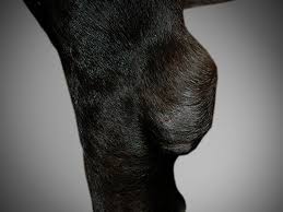
Hygromas are pouches full of serous fluid enclosed in a capsule of fibrous connective tissue around the olecranon area, in the elbow joint. In some cases they may also be present around the hock, although this is much less frequent. They are a reaction to repetitive trauma in the affected area, or a consequence of continuous pressure being exerted on these areas when the animal rests on hard surfaces. Therefore, it nearly always affects large or giant breeds of dog, such as Great Danes, St Bernards or Dalmatians, among others. This is due to the fact that their elbows have to support a larger amount of weight when they are resting. However, hygromas can also appear on the elbows of smaller breeds of dog. They appear in dogs that spend a lot of time resting, or dogs that have undergone surgery and therefore require a larger amount of rest, where the decubitus position is recommended. Below we will briefly explain the main treatment that is provided to dogs with hygromas. We will place particular emphasis on the work of the authors Pavletic and Brum (2015) that have proved that the active drainage system is a highly recommendable solution.
Various types of treatment are available. The conservative type consists of protecting the elbow joints using soft bedding or blankets that absorb the pressure that is exerted on the elbows. If the hygromas have appeared as a result of repetitive trauma, using joint protectors when the dog is at its most active is recommended as a preventative measure. Conservative techniques also include hygroma suction and injecting corticosteroids, although there are no conclusive results to show that the issue can be completely resolved using these techniques (Pavletic and Brum, 2015). Conservative surgical treatment consists of passive drainage using Penrose drainage tubing. However, the use of this type of drainage involves a risk of contamination from the outgoing liquid which is retained in the dressing that is in contact with the skin. The technique used involves securing the Penrose drainage tubing and protecting the area with a heavy bandage that is removed and replaced every 3 to 5 days, depending on whether this is necessary and also depending on how much liquid has accumulated in the cotton filling inside the dressing. This procedure will be repeated and can go on for more than 3-4 weeks. Then the drainage system is removed. The use of soft bedding is recommended in the future as a preventative method, as a relapse is very likely.
In order to discuss the active drainage technique, we would like to concentrate on the study presented by the aforementioned authors (Pavletic and Brum, 2015). They documented the case of a 1-year old St.Bernard that suffered from bilateral hygromas. One was on the left elbow (diameter of 8cm) and the other on the right elbow (diameter of 4cm). A surgical intervention was performed on the larger hygroma (the one on the left), using an active drainage system which was secured to the cervical collar of the dog. Oral antibiotics were also administered to the dog every 12 hours for 7 days, as well as tramadol, which was administered for 3 days. The owner was shown how to use and empty the active drainage system. Soft bedding was also provided for the dog to rest. When the drainage was being administered, a reduction in the dog’s activity was also recommended. The owner was monitoring the quantity of fluid in the deposit and its appearance every 24 hours. The dog was assessed every week for three weeks, until the date the drainage system was removed. Over the following 18 months there was no evidence of hygroma recurrence. However, the hygroma on the right elbow that was not treated using the active drainage system had increased slightly in size. Finally, permanent use of soft bedding was recommended.
The main advantage of a minimally invasive active drainage system as opposed to a passive drainage system is the fact that the risk of contamination is reduced, as the liquid that is drained off is stored directly in the deposit. This also means that you can assess how much fluid is going out of the hygroma, as well as its appearance and possible changes in the latter. Also, if the system is obstructed, it is possible to detect this quickly, which is more difficult in the case of passive drainage. The system is also easy to secure and maintain, and does not need the same level of precautions regarding poisoning to be taken as the other systems. Therefore, even though it is necessary to reduce the amount of physical activity an animal carries out for three weeks to one month, the animal can still maintain its normal routine, since the limb will not need to be immobilised. Conclusively, the system is also more economical than passive drainage using Penrose tubing or more aggressive surgical techniques.
*M.M. Pavletic and D.E. Brum. Successful closed suction drain management of a canine elbow hygroma. 2015. Journal of Small Animal Practice. 56, 476-479.
Clara Castells Urgell
Veterinarian
![]()