Physical therapy for a successful recovery
A shoulder’s joint is spheroidal allowing for a wide range of motion, mainly of flexion and extension but also of abduction and adduction. In this joint the humeral head articulates with the glenoid cavity of the scapula. Thus the stability depends on the set of structures that we group together as active mechanisms and passive mechanisms. The passive mechanisms are mostly composed of medial and lateral glenohumeral ligaments, the joint capsule, subscapularis ligament and the reduced joint liquid content. Regarding the active mechanisms, these are composed of the combination of the previously mentioned structures (passive mechanisms) together with the bicep muscles, supscapularis, infraspinatus and teres minor. When contracting, these help stabilize the shoulder joint as they place the humeral head within the glenoid. Below, we stress the important role of the muscles and, therefore, physical therapy for generating stability in this joint when it has been altered.
There is little reported about the etiology of shoulder instability and what is described 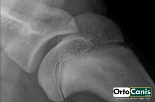 ranges from severe traumas to repeated microtraumas as factors that create this instability (Rials, 2015)1.
 To diagnose this condition, the following clinical signs must be present: First, the customer will inform us of a limp that their dog has had for a long time, although it may only be present after intense exercise. Second, in an examination under sedation, we will determine that there is also pain upon palpation and extension of the shoulder, as well as possible atrophy of a variable degree of the muscles that make up the shoulder. On the other hand, we will carry out craniocaudal stability tests and abduction/adduction tests, comparing the results with the contralateral limb. We will also perform radiological tests in normal conditions and under stress, although they will not always be of much use. Often they enable us to detect signs of osteoarthritis and, in some cases, signs of calcification of the bicep or supraspinatus tendons. Arthrocentesis will also help us with the diagnosis. With the sum of all these tests we will be able to determine if the process is intraarticular or periarticular. If it is intraarticular, we will explore the joint arthroscopically. (Trilla, 2005)2.
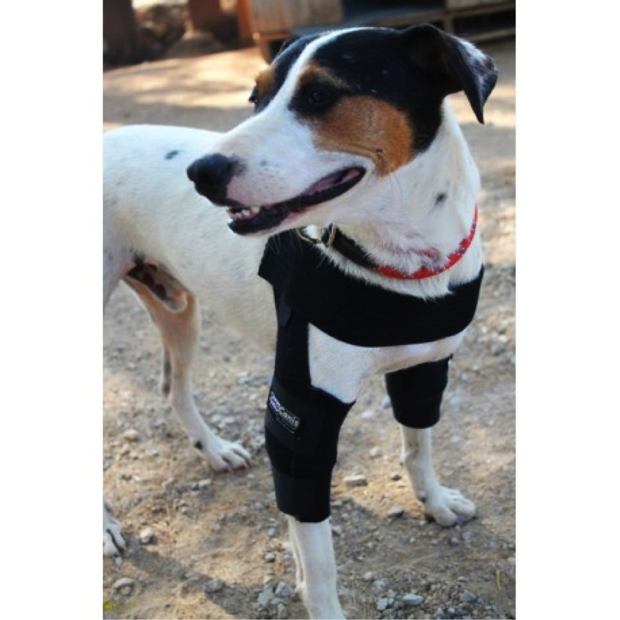
Treatment of shoulder instability can be done medically or surgically. The medical treatment consists of restricting movements and reducing pain. Natural anti-inflammatories can be helpful for this purpose if the veterinarian recommends administering anti-inflammatories for reducing pain. An elbow orthotic can also be a big help, which includes a double pectoral band that reduces the degree of abduction of the shoulder, where necessary. It is worth highlighting that in a study carried out by Rials published in the SEVC of AVEPA in 2013, from a total of 38 shoulders of the 35 patients that were studied, 7 were treated only with the conservative treatment and physical therapy for their recovery, which was complete and acceptable except for in just one case (that is, 85.8%). Therefore, the good results highlight the key role of the active mechanisms (the muscles) to create stability in this joint. With regards to the other 31 patients, they were treated with surgical techniques and physical therapy, with complete and acceptable recovery results in 88.6% of the cases, thus achieving a very similar percentage to those treated with the conservative treatment and physical therapy. The surgical techniques used were tenotomy of the biceps tendon in 3 cases and extracapsular tunnel suture in scapula and humerus anchor in 28 of the cases (Rials, 2013)3.
This study also demonstrated the importance of the medial glenohumeral ligament in the existence of shoulder instability, out of the 35 cases in the study only one had this structure unaffected. The integrity of this ligament is therefore crucial for achieving stability in the shoulder joint. Regarding the synovitis, it was found in 100% of the cases. The biceps tendinitis on the other hand was only found in 37.14% of the cases, the injury of the subscapular tendon in 42.56% and the labrum in 2.86%.
1 José manuel Rial Cels. Artoscopia del hombro. Estudio retrospectivo de 16 casos clínicos de inestabilidad de hombro. ARGOS 53/2015. Hospital veterinarios Marina Baixa. Spain.
2 Victor Trilla Muntanyola. Exploración artroscópìca del hombro en el perro. ARGOS 566/2005. Hospital Veterinari del Maresme. Spain.
3Jose manuel Rial Cels. Hallazgos artroscópicos en 35 perros con inestabilidad de hombro. Póster publicado en el SEVC de AVEPA el 2013. Hospital Veterinario Marina Baixa. Spain.
Ortocanis Veterinarian
![]()