There is talk of patellar dislocation in dogs when the kneecap, the small bone located in front of the knee joint whose correct positioning is necessary for proper functioning of the animal’s limb, is displaced from the trochlea, causing pain and functional weakness in the dog.
Medial luxation is the most common type of dislocation. It occurs in 80% of the cases while lateral only occurs in 20%. Approximately 30% to 50% of the cases are bilateral and it is more common in females than in males, especially in small and toy breeds.
Lateral dislocations can happen in small adult breeds and in 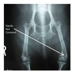 puppies of large and giant breeds.
This condition is characterized by a misalignment of the limb, deformations occur during the animal’s development which force the kneecap out of place. It can be caused by congenital disease or, in some cases, be due to trauma.
It is advised that dogs suffering from this congenital disease are not used for reproduction, as the condition is passed on through generations.
Some dogs suffer from a dislocated kneecap due to trauma. In these cases the dislocation is generally associated with a torn anterior cruciate ligament in the knee.
Depending on the clinical symptoms and subsequent radiological findings, dislocations can be classified in 4 degrees:
Degree I – Intermittent patella dislocation causing limpness in the limb when it is out of place. In the dynamic examination, the leg is lifted every three or four steps by bending the knee or with a small jump.
Degree II – Dislocation occurring more frequently than Degree I. The kneecap is easily dislocated. There is slight external rotation of the leg. Many dogs can live with this degree for many years prior to progressive arthritis that manifests with a limp or even more serious causes.
Degree III and IV – The kneecap is permanently dislocated, with a very noticeable external rotation of the leg. There is moderate lameness. When bilateral, dogs walk with bowed legs, turning their feet inwards and carrying the weight on their forelimbs. In more severe cases it can be confused with hip problems.
In addition, the animal has pain, crackles and heightened sensitivity of the knee, which may lead to a decrease in activity, even refusing to go up and down the stairs or on the sofa, or get in and out of the car.
Although treatment depends on the degree of luxation and lameness, most cases require surgical treatment that consists of repairing the soft tissues, bone reconstruction, or a combination of the two. There are an infinite number of techniques and the orthopedic veterinarian will choose the most appropriate for each case.
Among the most commonly used techniques are: Overlap of the medial or lateral retinaculum, overlap of the fascia lata, anti-rotational suturing of the patellar and tibia ligaments, desmotomy/capsulectomy, quadriceps release, trochleoplasty (trochlear chondroplasty, sulcoplasty resection, trochlear sulcoplasty), tibia tuberosity transposition, patellectomy, osteotomy ...
 A new conservative orthopedic technique for treating knee dislocation is using a Hinged Knee brace for dogs that keeps the patella within the femoral condyles in order to avoid pain and instability. These braces are custom made and are very useful in cases where surgery has failed, or for different reasons when an operation is impossible or not wanted.
Animal Physical Therapist
Collaborator Ortocanis.com
![]()