Blind people use a cane to orient themselves in space and know where there are obstacles. Other animals naturally use sound, whiskers or other warning devices.

Perro ciego

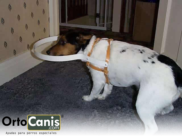
A blind dog also needs something to tell it when it is approaching an obstacle. Dennis Hacker, a veterinarian from California, told us how to manually make a “cane” for a blind dog.

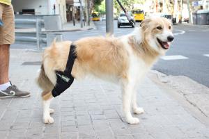
Using an aluminum strip, which allows to give it the desired shape while providing a solid barrier for the dog, we make a ring that fits the harness, with a prudent angle so that it can turn its head and have mobility in it.

Medidas de bastón para perro ciego

To manufacture it, we need to take 3 measures:

A – From the front leg to the neck.
B – From the neck to the tip of the nose.
C – The diameter of the chest just behind the front legs.


Necessary material:

A – An aluminum strip of the correct size, these are the measures A + B multiplied by 3.
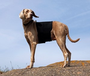
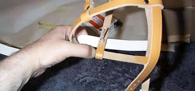
B – Leather harness, to fit on the chest. Remember that blind dogs tend to gain weight.
C – A riveting machine.
D – Rivets of 5 mm and 3-6 mm of reach.
E – Electric drill.
F – 5 mm helical drill bit.
G – Sandpaper.

The process for manufacturing is as follows:

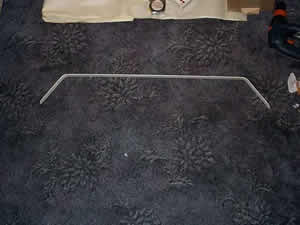
Sand the edges of the aluminum so that they are round.

lija1 lija2

Ortocanis.comAttach the harness to the blind dog and adjust it to make it comfortable. It measures the distance between the centers of the vertical bands. This is where the ring-shaped aluminum bar is going to be put.

Medir distancia de las bandas del arnés para el perro ciego arnes3

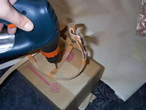
Drill four holes into the aluminum strip. A hole at each end and then another pair of holes at the distance between the two harness fasteners (as indicated in the photograph above left).

Medidas para la barra de aluminio del arnés

Pierce the harness at the bottom of the vertical strips of the neck where we measure. This is where the aluminum band is attached to warn the dog that it is about to hit itself.

taladro

Bend the distance to the ends of the aluminum bar with an angle of 45 degrees. Then, bend gently and in the opposite direction to give shape of circumference to the central area.

aluminio aluminio3

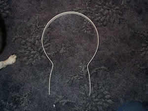
Rivet the aluminum ring to the harness on the vertical leather straps closest to the dog’s head.

Arnés perro ciego

Now, with the harness on the dog, level the hoop at eye and nose level. Mark the measurement and rivet the ring on the back of the harness.

Arnés para perro ciego

We already have our cane ready for blind dogs!

Arnés para perros invidentes

The result is phascinating, a solution that will improve the lives of dogs with vision problems.
At first they feel confused and need to adapt, as soon as they understand the operation and see the improvement they quickly dodge the obstacles.

In case you are not one of those who have a lot of patience, the new harness for blind dogs is now available for sale.

Arnés para perro discapacitado


The most common cause of femur fracture is a blow to the thigh bone area. Unless you have witnessed the accident firsthand, the first symptom or suspicion that the dog has broken its femur may be lameness. If the femur fracture is not repaired, the dog can have serious complications. Let’s explain how recognize and treat femur fracture in dogs.Femur y Tibia perro

  1. Dogs that suffer this injury may experience lameness in the injured paw or may become immobile because of the pain they are suffering.
  2. Take the dog to the vet and try to let them know of your arrival. Ask someone to help you get the dog into the car. This will prevent your dog from walking on the injured paw.
  3. The veterinarian will need to take x-rays to confirm if there is a fractured femur. It will also determine if you can give your pet painkillers to relieve the pains.
  4. He or she will likely recommend surgery to repair your femur fracture. Unlike other fractures, the femur stabilizes more successfully after surgery. This lesion cannot be stabilized with a splint or cast, so surgery should be seriously considered.
  5. Once back home the activity should be very limited. Reduce the walk when you take it out to relieve itself. Keep the area warm and prevent it from resting on hard or cold surfaces. This will aid in healing and prevent further damage.
  6. Colchón para perros en rehabilitaciónChoose a quiet place for the recupyour dog’s eration. Dogs in rehabilitation need a quiet place to heal. Let him rest in a good bed and hold his favorite toys. Encourage your pet to rest and remind visitors that the dog needs to rest.


Don’t try to stabilize the fractured femur at home. Some dog owners try to splint or plaster the limb at home. Always check with your veterinarian about the options available.

Arneses para perros con movilidad reducida

Physiotherapy as an alternative treatment in compression of Cauda Equina


SUMMARY

The cauda equina, is the set of nerves and nerve roots that originate from L6 – L7 innervating the posterior train by means of the nerves: Sciatic, Obturator, Pudendo, Femoral and Pelvic.

When generating a trauma at this level, mild to severe compressions can occur generating in the patient pain, claudication of hind limbs, contracture of all muscle mass, proprioceptive deficit, ataxic gait, incontinence, among other clinical signs that will guide the Veterinarian to establish an adequate medical treatment and in the case of the physiotherapist, locate the areas of pain and improve weight support, resistance, bodybuilding (avoiding muscle atrophy as much as possible), joint mobility and proprioception.

The diagnosis is made by radiological study and magnetic resonance imaging of the spine at the level of the lumbo-sacral joint, treatment options

consist of: cage rest, allopathic (NSAIDs, analgesics, corticosteroids), homotoxicological, physiotherapy and rehabilitation, being an excellent option the synergy between homotoxicological treatment and physiotherapy (integral analgesia).

This paper presents the case of a patient of a Pinscher, female, 10 months, which is taken to consultation after having suffered a trauma by fall presenting as main signs paraparesis, contracture of all muscle mass in hind limbs, proprioceptive deficit and ataxic gait. It was decided to perform physiotherapy and rehabilitation without suspending the medical treatment established from the beginning, managing to reduce the recovery time, improvement in the patient’s condition, elimination of the cause of the physical alteration, pain relief, reduce inflammation and resumption to daily life.

Keywords: cauda equine compression, physiotherapy and rehabilitation.

Interested? Here’s the full article


Angelica B. Ortega Vasquez

Ortocanis.com Collaborator


Hip dysplasia is a very common problem in certain breeds: BullDog, Bordeaux Doge, St. Bernard, Neapolitan Mastiff, German Shepherd, Rottweiler, Golden… all of them have an incidence above 20%.

Dysplasia is a multifactorial, multigenic and hereditary disease, that is, there are several factors that predispose and cause hip dysplasia, there are several genes involved in its appearance and it has a hereditary character.

Environmental factors are becoming more and more important in the development of hip dysplasia, the genetic factor is necessary to develop the disease but this is not the only factor. Genetics is a necessary but not exclusive factor, that is, you can have the genetic predisposition and not develop the disease but if you do not have a predisposition it is sure that it does not develop.

There are several degrees of dysplasia, and also those that appear when the dog is a puppy or those that give problem already in adulthood; but in this article we will focus on the treatment and specifically on the treatment of dysplasia to young dogs.

Classification of the degrees of dysplasia according to the OFA :

Grade I: minimal alteration with small subluxation and few degenerative changes.

Grade II: marked lateral subluxation of the femoral head, 25-50% of which is outside the acetabulum.

Grade III: 50-75% of the femoral head is outside the acetabulum; there are important degenerative changes.

Grade IV: dislocation of the femoral head with flattening of the acetabular border and femoral head; there are major degenerative changes.

The presentation in the young dog is puppy hip x-ray normally between 5 and 6 months and is marked by a significant limp.

A dog is not considered to be free of dysplasia until at two years of age already completed no problems or inconsistencies are observed in the control x-rays.

Food is one of the factors that predisposes to the appearance of hip dysplasia, Calcium-Phosphorus imbalances that must keep a correlation Ca1.6% – P1.1% and above all not overfeeding or providing excess proteins allows us to minimize the incidence of hip dysplasia. A hypocaloric diet from 3 months to 8 months protects dogs with rapid growth from dysplasia. Excess weight at 60 days is another factor that predisposes to the disease.

Hip dysplasia in the puppy usually debuts from 5 or 6 months, before it is not possible to observe any problem and the dog has been completely normal and has developed normally. The debut is usually presented as a sharp limp that prevents the dog from playing as it had done to date. We can observe changes in the desire to play, negative when going for a walk, to relate to other dogs or owners. Mood swings, frequent slips of the hind legs, discomfort and even refusal to be touched and the fact of “fleeing” from children in dogs that until a few days ago were playful and affectionate are frequent.

Sometimes when you reach 90% of the growth between 8 and 11 months the signs can be reduced and even disappear. Anyway the dysplasia remains and in many cases the problems reappear after a while and sooner rather than later signs of osteoarthritis appear in the hips.

The most common clinical signs are:

Lameness that may increase with exercise

Walking and jogging with hip swing

Morning stiffness

Difficulty getting up

Muscle atrophy

Refusal to move

Mood swings

Pain on palpation

Sign of Ortolani.

Although there are surgical methods: excision of the pectineus muscle, triple hip osteotomy, arthroplasty of the femoral head, osteotomy of the pubis, forage, hip prostheses most are practiced when the dog is young to supposedly decrease the possibility of secondary coxofemoral osteoarthritis in adulthood. The hip prosthesis should be reserved for severe cases and once the growth has finished.

Medical treatment is based on anti-inflammatories, we can start with natural anti-inflammatories, such as inflamex, which does not contain medicinal substances, if we do not obtain the expected results move to Aine’s and in extreme cases corticos are resorted to. We must include nutraceuticals especially chondroprotectors since they reduce the incidence of osteoarthritis and protect the articular cartilage. These are used in senior dogs in a very general way but are very useful as a joint protector in growing dogs, there are specific drug carriers for young dogs. Weight reduction, moderate and above all regular exercise are other basic points, as well as improvements in the environment and the fact of sleeping on a special mattress for older dogs and in a warm place away from humidity.

Canine physiotherapy can help a lot to better develop the muscles to reduce pain, to draw tensions and eliminate compensations that the dog has made with the wrong postures and antialgic positions. This will be based on TENS, ultrasound, therapeutic exercises, the use of hydrotherapy, laser, shock waves…

The main improvement in the environment is to sleep on a good therapeutic mattress, not to be cold or exposed to a lot of humidity, to use in winter a thermal coat for dogs, to be able to be a therapeutic canine blanket that can be used all year round.

We can help our Dog with hip dysplasia, regular physical exercise can be very useful to improve muscle mass that better withstands poor joint congruence, avoid impacts, jumps or uncontrolled runs during the presentation of the picture are also important elements. Physiotherapy and massages allow you to always have the dog in a correct muscular state, and all the adjuvant treatments such as acupuncture, massages, reiki, bach flowers … they can also help with treatment. The latest novelty is the hip supports that help stabilize the pelvis, give support and greatly improve the quality of life of our pets.

Toni

Ortocanis.com Team

Deforming Spondylosis in Dogs

DEGENERATION OF THE SPINE IN DOGS

Deforming spondylosis is a degenerative, non-inflammatory disease of the spine, characterized by the production of bone spurs in the lower, sides, and upper area of the vertebrae of the spine. These spurs are simply caused by bone growths, usually growing in response to aging or injury.

In dogs, deforming spondylosis most often occurs along the spine, in the back of the chest, and in the upper vertebrae of the lower back. Older and large breed dogs are at higher risk of developing deforming spondylosis.

SYMPTOMS

*Patients are usually asymptomatic, bone growth can be felt by touching your pet before perceiving changes in its behavior in the wake of growth

  • Fracture of spurs or bridges can cause pain
  • Rigidity
  • Restricted movement
  • Pain

CAUSES

Repeated microtrauma – repeated pressure on the same joints or bones, through certain exercises or activities
Major traumas – the body responds when trying to grow new bone
*Predisposition to hered spurs

DIAGNOSIS

Your veterinarian will do a complete physical examination of your dog, including a biochemical profile, a complete blood count, a urinalysis, and an electrolyte panel, in order to rule out or confirm other diseases, such as cancer. You will need to give a complete history of your dog’s health, including background history of symptoms, onset of symptoms, and possible incidents that may have precipitated this condition.
X-ray images of the chest and abdomen (side view) are essential for the diagnosis of deforming spondylosis. X-rays reveal osteophytes (small bone growths) in the vertebrae, or in more advanced cases an osteophyte can be found as a bridge in the space between the vertebrae.

Your doctor may choose from several other types of tests in order to reach a definitive conclusion. A myelography uses the injection of a radiopaque substance to obtain an interior image; Computed tomography (CT) or magnetic resonance imaging (MRI) are also options. These procedures can help your veterinarian find a bone spur that may be putting pressure on your dog’s spinal cord or nerves (leading to neurological reactions).

TREATMENT

faja para el dolor de la espalda de perroUsually, patients with deforming spondylosis do not show abnormal external symptoms of initial bone growth. A neurological examination should be performed to rule out a pathology of the spine that requires surgery. Otherwise, if the growth has reached the point of damaging nerves and tissue, and your pet suffers intense pain, or if your veterinarian has decided on a surgical solution, your dog will be hospitalized. Under normal circumstances, when damage to the body is minimal, and your dog feels little discomfort and pain, the condition will be treated on an outpatient basis, with strict rest and prescription painkillers for home treatment. Pain medications are given after your meals. To speed up recovery and from four days after the intervention you can use thermal coats or spine girdles for dogs (consult your veterinarian). Acupuncture can also provide pain relief for some animals.

LIFE AND MANAGEMENT

Depending on the severity of the symptoms, your veterinarian will schedule checkups to track your dog’s progress. Only give pain medication when your dog is showing signs of discomfort (after a meal), and only in the exact amount prescribed, unless your veterinarian tells you otherwise. Drug or drug overdose is one of the most common causes of unintentional deaths in pets. You will need to provide a safe and quiet place for your dog to rest, away from other pets and active children. During this time, limit yourself to slow walks around the neighborhood. When your dog shows no signs of discomfort for several weeks he can slowly return to normal activity.

Source: Venfido
www.ortocanis.com

What is the proprioceptive system?

It is the system by which the brain receives information about the position and movement of body parts with each other and in relation to their support base. This is produced through a series of receptors distributed throughout the body. Proprioceptive sensitivity is extraordinarily important in the dog’s relationship life.

Within the physiotherapy examination (musculoskeletal or neurological), the proprioceptive system will always be evaluated to have a reference and guidelines with which to establish the treatment plan. Sometimes the same techniques that help us to value the animal, serve us later to rehabilitate it.

Postural reactions

  1. Positional reaction or conscious proprioception
    1. Put the dorsal face of the limb in contact with the ground. The animal should instantly rectify to the normal position.
    2. Put the limb of the animal in abduction or adduction. In this case you should also instantly rectify towards the starting position.
    3. Place a sheet of paper so that the dog supports the pads. Move the leaf laterally in thoracic limbs and caudolaterally for the pelvic limbs. When perceiving the movement stimulus, you need to reposition the limb correctly.
  2. Hemistation
    1. You should take the limbs of a hemibody and move the animal to the opposite side to see if it is able to withstand the balance.
    2. In the case of musculoskeletal injuries it is also possible to assess the affected limb by taking only the opposite limb or one of the previous ones and assess the balance reaction presented by the animal (uniestación)
  3. Hemimarcha
    1. It is exactly the same exercise as the hemistation only that the animal will have to move on the two limbs.
  4. Jump test
    1. Holding the animal and preventing it from supporting three of its four limbs, move it laterally. You will need to make small jumps.
  5. Truck test
    1. Make him move with his thoracic limbs holding him through the pelvic area. Do it with the head of the animal in a high position to prevent it from looking where to put your hands.
  6. Extensor postural impulse reaction test
    1. Take the animal by the armpits in an upright position and descend it slowly. When its hind limbs touch the ground, it will extend them caudally in a marching motion, anticipating displacement.
  7. Visual and tactile reaction
    1. Holding the animal, he approaches the edge of a table until he touches it. You should place your limb on the table instantly. We will assess visual and proprioceptive sensitivity. It should be repeated by covering the eyes, so we will value tactile and proprioceptive sensitivity.

Balance in season and dynamics

The balance of the animal can be assessed by provoking some of the postural reactions mentioned above or by creating destabilizations of the animal in static and during walking.

During walking you can tap small sideways to see how it reacts. If the animal has its correct balance, it will continue walking in a straight line without modifying its state or its rhythm.

Coordination

Coordination can be assessed through circles, eights, climbs, descents, speed changes, different terrains with small difficulties in between.


Ortocanis Writing

[email protected]

A dog’s ability to move and perform its daily activities depends on the ability of the brain, spine, nerves, and muscles to coordinate together.

This complex communication system exchanges information when nerves in the brain send messages to the body about the outside environment, and the body sends messages to the brain about what it is experiencing in the environment. These messages are transmitted through nerves in the spinal cord, which are embedded in the spine or spine. Together, the nerves of the brain and spinal cord make up the central nervous system. Trauma to any part of the nerve pathways can lead to poor communication, or a total lack of communication between the brain and the body, leading to an inability to coordinate body movements.

Silla de ruedas para perro

The spine is made up of a set of 24 bones called vertebrae, which are separated from each other by small cushions called intervertebral discs. Together, the vertebrae and intervertebral discs protect the spine from damage. Trauma to the vertebrae or discs can create vulnerability in the nerves of the spinal cord, leading to new disorders to the neural pathway.

When a dog experiences paralysis, it is often because communication between the spinal cord and the brain has been disrupted. In some cases, the dog is not able to move its legs, a state of total paralysis, and in other cases, there may still be some communication between the brain and the spine, and the dog will only appear weak or have difficulty moving its legs, a condition known as: partial paralysis. There are also cases where a dog may be paralyzed on all four legs (quadriplegia), and in others, the dog may be able to control movement in some of its legs, but not all. This is determined by the location of the trauma, occurring in the brain, spine, nerves or muscles.

Some breeds are more prone than others. Dogs that are close to the ground with elongated backs, such as dachshunds and bassets, are commonly affected by ruptured vertebral discs by putting pressure on the spinal cord, a condition known as intervertebral disc disease. Some breeds are genetically predisposed to a condition called degenerative myelopathy (MD), a disease that attacks nerves on the backs of older dogs (more than seven years old). It is a slow action, a progressive disorder that eventually leads to paralysis of the hind legs. Breeds affected by this disease are the Welsh corgi, boxer, German shepherd, Chesapeake Bay retriever, and Irish setter.

Symptoms:

  • Not able to move all four legs (quadriplegia)
  • Not able to move the hind legs (paraplegia)
  • Walks with front legs while dragging hind legs
  • There may be pain in the neck, spine, or legs
  • Can’t urinate
  • Not able to control urination, urine dripping
  • Not able to control defecation
  • Constipation

Causes:

  • Canine degenerative myelopathy (MD) – related to the gene of German shepherd, boxer, Welsh corgi, Chesapeake Bay retriever, ages 7-14 years; the cause is unknown
  • Slippage of the discs in the back – intervertebral disc disease
  • Spondylodiscitis – bacterial or viral infection in the bones of the spine (vertebrae)
  • Infection or inflammation of the spine
  • Distemper
  • Meningomyelitis – viral or bacterial infection of the brain, leading to poor communication of nerve impulses
  • Polymyositis – infection or inflammation in the muscles
  • Polyneuritis – inflammation of the nerves
  • Embolism – obstruction of blood flow to the spine
  • Aortic embolism – obstruction of blood flow to the back legs
  • Tumors or cancer in the spine or brain
  • Tick paralysis as a result of tick bites
  • Rocky Mountain Fever
  • Botulism – bacterial toxins
  • Myasthenia Gravis – severe muscle weakness
  • Fibrocartilaginous embolism the fluid inside an injured disc enters the arterial system and settles in the spinal cord, creating a permanent embolism, or blockage, is irreversible, but not progressive.
  • Hypothyroidism – Low thyroid level
  • Spinal injury
  • Malformation of the spine or vertebrae

Diagnosis:

You will need to give a complete history of your dog’s health, the onset of symptoms and possible incidences that may have led to this condition, such as tick bites, or recent injuries from jumping or falling. During the physical exam, your veterinarian will pay close attention to how well your dog is able to move his legs, and how well he is able to respond to reflex tests. The veterinarian will also test your dog’s ability to feel pain on all four legs, control of the head, spine, and legs for signs of pain and alertness to the touch.Perro atropellado y rescatado por perrera de barcelona

All of these things will help your veterinarian locate the place in your dog’s spine, where nerves or muscles are having problems. Basic laboratory tests include a complete blood count, a biochemical profile and a urinalysis, which can determine if your dog has a bacterial infection, a virus or a toxin reaction, which is interfering with nerve pathways. X-ray images of the spine can show evidence of an infection, a malformation of the vertebrae, or a herniated disc that is pressing against the spinal cord. Other conditions that can lead to disruption of nerve pathways may be evident on an X-ray, such as tumors, obstructions, or inflamed nerves.

In some cases, your veterinarian may order a special X-ray called a myelogram. This process uses an injection with a contrast agent (dye) into the spine, followed by X-ray images that will allow the doctor to see the spinal cord and vertebrae in more detail. If these imaging techniques aren’t helpful, your veterinarian may order a computed tomography (CT) scan, or magnetic resonance imaging (MRI) of your dog’s brain and spine, both of which provide a very detailed picture. In some cases, your veterinarian may take a sample of the fluid around the spine, for analysis, or sample of the muscles or nerve fibers for a biopsy. These tests can determine the presence of an infection in the brain or spine.

Treatment:

The course of treatment will depend on the cause of the paralysis. If your dog is unable to walk, urinate, or defecate on his own, he will most likely be admitted to the hospital while his veterinarian will work to deliver a diagnosis. From there your veterinarian will track your dog’s recovery and progress. If your dog is in pain, he will be given medication to help control the pain, the bladder is emptied several times a day by catheter, and your dog will be physically moved to make sure he doesn’t get ulcers from staying in one place for too long. There are several products that will help you spend time still without getting damaged, such as mattresses. If the cause of the paralysis is an infection or a herniated disc, the condition will be treated with either medication, surgery, or therapy. Anti-inflammatory drugs that may be natural and specific such as imflamex will be used to reduce inflammation of the nerves. Tumors or blockages of blood flow can be repaired surgically, depending on the vulnerability of the location. Some paralyzed dogs recover very quickly. Depending on the severity of the illness, your dog may stay in the hospital until he is able to walk, or your veterinarian will decide to send your dog home with a guide to provide care and recovery at home. Your veterinarian will establish a plan to review the progress of your dog’s treatment and adjust it accordingly.

life and a lot!

perro-rodilla-en-la-calleYour veterinarian will help you make a plan for your dog’s care at home. Occasionally, the dog may resist care due to pain, but assertive and gentle care will help decrease fear reactions.

It is important that you take care of your dog properly so that he can fully recover. Carefully follow all instructions from your veterinarian. If your veterinarian has prescribed medication, be sure to administer all treatment, even after your dog appears to have fully recovered. In most cases, a canine wheelchair can be of great help, either for the rehabilitation process or as a simple means of transporting the immobile part of the animal. Most wheelchair dogs adapt very well and continue to enjoy their life.

Orthocanis

Ayudas para perros con artrosis

source:VenFido

Although genetics is the determining part of dysplasia by 99% percent, it is nutrition that is the most important part. Once you have diagnosed dysplasia you can NOT do anything genetically, you only have to influence nutrition and physiotherapy. It’s that simple.

I teach you five key secrets in nutrition to make your best amig@’s dysplasia more bearable. In addition to an Expert in Canine Nutrition, do not forget to also consult one in canine physiotherapy, it can help you a lot.

Nutrición Ortocanis

GRAINS IN THE DIET

Unfortunately the vast majority of commercial veterinary diets for dogs are bad, why? Because they have a lot of carbohydrates in the form of grains and / or cereals, they do it to reduce costs, they are cheaper than animal protein, the nutrient that your dog really needs.

Dietary grains or cereals have been shown in dogs to promote continuous and exaggerated insulin secretion* as well as joint inflammation. I remind you that your dog does not have the physiology to split starches, carbohydrates, that is, grains and cereals. It is a carnivore, do not forget, do not feed it as if it were cow or chicken.

GLYCOSAMINOGLYCANS

Don’t panic with the name. They are nutrients that promote cartilage health. Patients with dysplasia problems (and arthritis in general) have shown that they can absorb toxic compounds or substances, some of which affect the joints.

How can we lessen this? If you give your perr@ some cartilage (rich in glycosaminoglycans) in the diet, these are poorly absorbed and remain in the intestinal lumen.

Glycosaminoglycans are complex carbohydrates that have the ability to adhere some of these toxic substances on their surface while they are in the lumen of the intestine, and thus be excreted in the excrement without passing into the bloodstream and therefore prevent their arrival and implantation in the joints.

A substitute for cartilage? There are nutritional supplements based on glycosaminoglycans, in short, they are more practical and come concentrated in a pill. There are many brands on the market: Cosequin, Synoquin, Hyal oral…

BELOW OR ABOVE WEIGHT?

If I have to choose to be slightly above my weight, I prefer the latter, and the same applies to my dog.

As an Expert in Canine Nutrition, this topic, that of weight, is one of the most recurrent. Owners of puppies of large and/or molossian breeds (mastiffs, Rottweiler, Siberians, shepherds, doges…) want “stuffed” puppies growing as much as possible. Gross error.

Remember, if you want a healthy dog orthopedically weight is key (it also applies to us) Have you seen overweight wolves, lions or hyenas in the wild?

How do you know if your dog is at an appropriate weight? The best allies for this are sight and touch. I recommend you visit the following link so that you learn how to determine the body condition of your dog.

BONES IN THE DIET

There are many myths in dog nutrition, created, above all, by the pet food industry. I remind you that your dog is a carnivore, every carnivore eats bones in nature, moose, rabbits, all kinds of birds (such as chicken or chicken) and ALL BONES, absolutely all, splinter and do not die!

It refreshes your memory. Commercial foods (pellets) have been massively and widespread in the last two decades. Before everyone gave homemade food (leftovers) including bones.

Bone is a mine of vitamins, fats and minerals of the highest quality, do not forget that bone is mainly made up of minerals such as calcium, phosphorus, fluoride, magnesium … water, and organic matter such as collagen. All of these nutrients are key to bones and joints, among other organs.

By the above I do not mean that you start introducing bones into the diet without knowing. COOKED BONES are the dangerous ones. The best diets are homemade, and if they include bones (RAW) occasionally, better.

NUTRITIONAL SUPPLEMENTS

If I am given the choice between providing a quality diet for a dog without supplements (the best diets are well-made homemade ones) or a poor quality diet and a quality supplement, I stay with the first without a doubt.

Now, if I can choose for my dog a quality homemade diet, alternated with a commercial diet, also of quality, and added with a specific supplement for his dysplasia problems Bingo!

With the arrival of orthomolecular nutrition (specific nutrients for specific situations) and the advancement of science, many nutrients appear that at the molecular level have a positive impact on the patient with dysplasia problems.

From vitamin E, bioflavonoids to omega three and enzymes. There are more and more of these nutrients for multiple situations. Regarding the subject we are dealing with, dysplasia, it would be something that you should not overlook and ask for advice from a specialist in the subject.

Carlos Alberto Gutierrez / Veterinarian collaborating with Ortocanis.com

Treatment of hygroma of the elbow

Elbow hygroma is a disease that mainly affects large short-haired dogs, such as Big Danes, Greyhounds and Dalmatians. In dogs with elbow hygroma, a fluid-filled sac appears on one or both of the dogs’ elbows. In rarer cases, hygromas can develop in the hock. In many cases, elbow hygromas do not cause pain or difficulty. However, they can become infected, which can be painful for the dog and requires treatment. Other animals susceptible to developing hygromas are dogs that are not very active or are recovering from illness or injury.

Causes
Hygromas develop because of recurrent trauma to a dog’s elbow or elbow. For example, resting constantly on wood, cement, or other hard surfaces can cause stress on the joint and cause hygroma. This condition mainly affects large dogs, as there is a greater weight on their elbow joints when they are lying down.Hygroma codo

Bandage
In some cases, bandaging a dog’s elbows can help prevent hygromas from getting worse. Bandages will prevent contact with hard surfaces and in turn avoid problems caused by hygroma. In addition, some products such as the canine elbow protector or the dog knee protector will prevent the hygroma from having additional contact with the hard ground. Ask your veterinarian how you can help your dog.

Drainage
Some veterinarians recommend that hygroma fluid be drained with a needle or syringe. Getting the fluid out is not considered an effective treatment because the needle can cause an infection. In addition, drainage can only improve the condition temporarily. If the dog continues to lie on hard surfaces, the hygroma will become larger, that is, the drainage will have to be done on a regular basis.

Surgery
Some veterinarians may recommend surgery to treat elbow hygroma, especially in cases where there is infection or when the hygroma has ulcerated. In surgery, the skin must be drained and removed. Since hygromas can grow quite large, it may be necessary to make skin grafts to cover the affected area. Healing from surgery takes about a month, and the dog will need to wear a splint during recovery to protect the affected area.

Prevention
The best way to prevent your dog from developing an elbow hygroma, or to prevent an existing one from worsening, is to provide the dog with a soft surface for him to rest and sleep. If there is no carpeted area available, make the dog a soft bed and leave it where he likes to lie down. In the event that the dog is inactive because he is recovering from an illness or injury, have him get up and move (at least enough to change the position he is lying in) several times a day.

Written by Anna Aronson

source: eHow

Canine osteoarthritis is a very common disease in dogs. It appears as a result of the inevitable evolution of a joint that ages or becomes increasingly fragile due to trauma or malformation. This is a very painful condition that needs to be treated right away. It can affect all the joints of the body, both those found in the anterior and posterior extremities, as well as those that form the spine. In the case of senior dogs, the most common is that this disorder affects several joints at once.

protector rodilla para perroThe articular surface is covered by a tissue called cartilage, which plays a role very similar to the shock absorbers found in cars. In addition, it prevents the underlying bone from deteriorating due to the repeated rubbing to which it is subjected by continuous movement. Osteoarthritis is characterized by a progressive destruction of this cartilage and by an abnormal bone proliferation at the edge of the articular surfaces known as osteophyte, also called ‘parrot beaks’ when they are located in the spine. The affected joints lose elasticity, cause pain and prevent the animal from moving normally.

Evolution of deterioration
As a rule, this ailment affects, first of all, the high joints of the limbs: hips and knees, shoulders and elbows. Symptoms are more or less important depending on the number of joints affected. However, there is an unequivocal sign that makes us intuit that the animal is affected. Lameness usually manifests itself when the dog gets up and starts up after remaining immobile for a long time.

The pain prompts the animal to avoid support on the affected limb and, being impeded, stops running, and of course, jumping. As it evolves, the pain increases. When making certain movements, the dog emits small moans, it is even possible that the animal is irascible and tends to present aggressiveness when we try to manipulate the affected joint.

In more advanced cases, the joint may be partially blocked, making it impossible to perform certain movements. At this point, the animal hesitates to use the diseased limb. In addition, this lack of activity leads to a significant deterioration of the musculature surrounding the joint. As a result, the diseased area begins to atrophy, which increasingly complicates its use.

Two fundamental types
As a general rule we distinguish two types of osteoarthritis: primary and secondary. The first type usually affects elderly animals and appears due to the normal aging suffered by the joints due to the passage of time. In fact, it is a progressive and inevitable wear of the articular cartilages. Usually, this type of osteoarthritis affects different points at the same time.

As far as secondary canine osteoarthritis is concerned, it appears as a consequence of a triggering factor, which causes the affected joint to stop functioning normally. For example, this type of osteoarthritis can appear due to trauma – a sprain, a fracture, etc. – or due to a birth malformation, such as hip dysplasia.

Another very common cause that causes the appearance of secondary osteoarthritis is obesity. If you do not control the diet your friend can have a weight well above the average that we find in the breed. The joints are not made to support such a significant overload of kilos, so they deteriorate easily. Unlike primary osteoarthritis, secondary osteoarthritis can affect animals of all ages and, as a general rule, usually affects only one joint.

Issue a diagnosis
The diagnosis of this disease can be based on three factors: the pathological history of the animal, gait examination and manipulation. When studying the history, the veterinarian must take into account old joint fractures, injuries that the animal has suffered a long time ago, as well as possible sprains. When observing the gait will look at whether the dog limps, even if only very slightly and incipiently, since lameness is an unequivocal symptom of the disease. Finally, when manipulating the affected area it is very possible for the animal to show signs of pain.

Often, the region in which the diseased joint is located is usually somewhat deformed due to osteophytes and muscle atrophy caused by the absence of physical activity. Many times a characteristic snap is detected when we move it. Through the radiological study, the animal health specialist will be able to determine the severity of osteoarthritis and establish the most appropriate treatment. Among the possible treatments, the appearance of specialized supports for some limbs that can effectively combat the deterioration of the animal’s quality of life stands out. The use of these supports is effective and has proven in countries such as the USA to be treatments as innovative as successful, however their use should be consulted with the veterinarian or animal physiotherapist.

Orthocanis Team